Tuesday Poster Session
Category: Colon
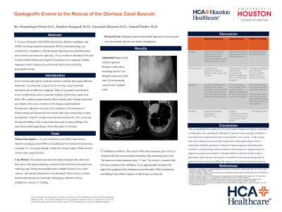
P3721 - Gastografin Enema to the Rescue of the Glorious Cecal Bascule
Tuesday, October 29, 2024
10:30 AM - 4:00 PM ET
Location: Exhibit Hall E

Has Audio
- BB
Bushra A. Bangash, MD
HCA Kingwood
Spring, TX
Presenting Author(s)
Dominique Pean, MD1, Bushra A. Bangash, MD2, Guntash Khasria, MD1, Ismail Hader, MD1, Ammar Hasan, MD1, Asad Rehman, DO1, Mahnoor Hashmi, MD1
1HCA Kingwood, Kingwood, TX; 2HCA Kingwood, Spring, TX
Introduction: Cecal bascule is the rarest type of cecal volvulus which involves the upward folding of the cecum without any twisting or torsion. This is thought to result most commonly from a large mobile cecum or postoperative adhesions. Delays in treatment can lead to severe complications such as ischemia, perforation, sepsis, and death. Treatment is usually surgical resection or cecopexy. Here we present a case of cecal bascule successfully treated with a gastrografin enema.
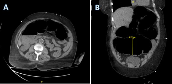
Case Description/Methods: A 74-year-old female with GERD, hiatal hernia, Barrett's esophagus, and COPD was hospitalized for treatment of pneumonia requiring 25 L of oxygen through a High Flow Nasal Canula. The patient reported lower abdominal pain and that her last bowel movement was eight days prior. Physical examination showed a distended abdomen with hypoactive bowel sounds. X-ray imaging revealed extensive gaseous dilatation of the colon, measuring up to 8.5 cm along the transverse colon and 12.8 cm along the cecum. During the hospitalization, multiple laxatives, two water enemas, and manual disimpaction was attempted without success. Gastroenterology was consulted for constipation and Surgery was consulted for possible large bowel obstruction. A CT scan of the abdomen and pelvis was done which showed that the cecum was located in the left lower quadrant of the abdomen, whereas it was previously appropriately located in the right lower quadrant in 2022. The abnormal cecal positioning was indicative of cecal bascule. A gastrografin enema was ordered to evaluate the large bowel and following this patient was able to have a large bowel movement and her symptoms resolved.
Discussion: This case highlights the complexity of Colonic Pseudo-Obstruction evolving into a rare cecal bascule. Cecal bascule remains the rarest type of cecal volvulus accounting for only 20% of all the cases. We present a rare case where it presented in a patient without any previous surgical history and resolved on its own with a gastrografin enema. Prompt recognition and treatment are essential to managing cecal bascules. Although definitive treatments include surgical approaches, more literature and research are needed for non-surgical management in patients who are not good candidates for surgery or who decline surgical interventions.

Note: The table for this abstract can be viewed in the ePoster Gallery section of the ACG 2024 ePoster Site or in The American Journal of Gastroenterology's abstract supplement issue, both of which will be available starting October 27, 2024.
Disclosures:
Dominique Pean, MD1, Bushra A. Bangash, MD2, Guntash Khasria, MD1, Ismail Hader, MD1, Ammar Hasan, MD1, Asad Rehman, DO1, Mahnoor Hashmi, MD1. P3721 - Gastografin Enema to the Rescue of the Glorious Cecal Bascule, ACG 2024 Annual Scientific Meeting Abstracts. Philadelphia, PA: American College of Gastroenterology.
1HCA Kingwood, Kingwood, TX; 2HCA Kingwood, Spring, TX
Introduction: Cecal bascule is the rarest type of cecal volvulus which involves the upward folding of the cecum without any twisting or torsion. This is thought to result most commonly from a large mobile cecum or postoperative adhesions. Delays in treatment can lead to severe complications such as ischemia, perforation, sepsis, and death. Treatment is usually surgical resection or cecopexy. Here we present a case of cecal bascule successfully treated with a gastrografin enema.
Case Description/Methods: A 74-year-old female with GERD, hiatal hernia, Barrett's esophagus, and COPD was hospitalized for treatment of pneumonia requiring 25 L of oxygen through a High Flow Nasal Canula. The patient reported lower abdominal pain and that her last bowel movement was eight days prior. Physical examination showed a distended abdomen with hypoactive bowel sounds. X-ray imaging revealed extensive gaseous dilatation of the colon, measuring up to 8.5 cm along the transverse colon and 12.8 cm along the cecum. During the hospitalization, multiple laxatives, two water enemas, and manual disimpaction was attempted without success. Gastroenterology was consulted for constipation and Surgery was consulted for possible large bowel obstruction. A CT scan of the abdomen and pelvis was done which showed that the cecum was located in the left lower quadrant of the abdomen, whereas it was previously appropriately located in the right lower quadrant in 2022. The abnormal cecal positioning was indicative of cecal bascule. A gastrografin enema was ordered to evaluate the large bowel and following this patient was able to have a large bowel movement and her symptoms resolved.
Discussion: This case highlights the complexity of Colonic Pseudo-Obstruction evolving into a rare cecal bascule. Cecal bascule remains the rarest type of cecal volvulus accounting for only 20% of all the cases. We present a rare case where it presented in a patient without any previous surgical history and resolved on its own with a gastrografin enema. Prompt recognition and treatment are essential to managing cecal bascules. Although definitive treatments include surgical approaches, more literature and research are needed for non-surgical management in patients who are not good candidates for surgery or who decline surgical interventions.

Figure: CT Abdomen and Pelvis: The cecum of the colon measures up to 10 cm in diameter with the more proximal ascending colon measuring up to 8 cm.
The transverse colon measures up to 7.7 mm. The cecum is located in the left lower quadrant of the abdomen.
The transverse colon measures up to 7.7 mm. The cecum is located in the left lower quadrant of the abdomen.
Note: The table for this abstract can be viewed in the ePoster Gallery section of the ACG 2024 ePoster Site or in The American Journal of Gastroenterology's abstract supplement issue, both of which will be available starting October 27, 2024.
Disclosures:
Dominique Pean indicated no relevant financial relationships.
Bushra Bangash indicated no relevant financial relationships.
Guntash Khasria indicated no relevant financial relationships.
Ismail Hader indicated no relevant financial relationships.
Ammar Hasan indicated no relevant financial relationships.
Asad Rehman indicated no relevant financial relationships.
Mahnoor Hashmi indicated no relevant financial relationships.
Dominique Pean, MD1, Bushra A. Bangash, MD2, Guntash Khasria, MD1, Ismail Hader, MD1, Ammar Hasan, MD1, Asad Rehman, DO1, Mahnoor Hashmi, MD1. P3721 - Gastografin Enema to the Rescue of the Glorious Cecal Bascule, ACG 2024 Annual Scientific Meeting Abstracts. Philadelphia, PA: American College of Gastroenterology.
