Tuesday Poster Session
Category: Colon
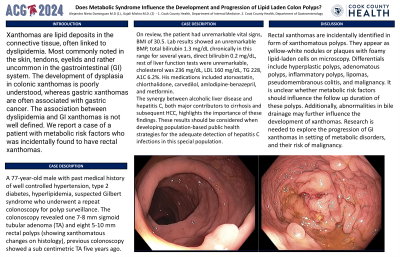
P3673 - Does Metabolic Syndrome Influence the Development and Progression of Lipid Laden Colon Polyps?
Tuesday, October 29, 2024
10:30 AM - 4:00 PM ET
Location: Exhibit Hall E


Alejandro J. Nieto Dominguez, MD
John H. Stroger, Jr. Hospital of Cook County
Chicago, IL
Presenting Author(s)
Alejandro J. Nieto Dominguez, MD1, Kajali Mishra, MD2
1John H. Stroger, Jr. Hospital of Cook County, Chicago, IL; 2Cook County Health and Hospital Systems, Chicago, IL
Introduction: Xanthomas are lipid deposits in the connective tissue, often linked to dyslipidemia. Most commonly noted in the skin, tendons, eyelids and rather uncommon in the gastrointestinal (GI) system. The development of dysplasia in colonic xanthomas is poorly understood, whereas gastric xanthomas are often associated with gastric cancer. The association between dyslipidemia and GI xanthomas is not well defined. We report a case of a patient with metabolic risk factors who was incidentally found to have rectal xanthomas.
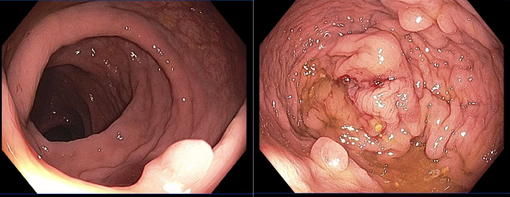
Case Description/Methods: A 77-year-old male with past medical history of well controlled hypertension, type 2 diabetes, hyperlipidemia, suspected Gilbert syndrome who underwent a repeat colonoscopy for polyp surveillance. The colonoscopy revealed one 7-8 mm sigmoid tubular adenoma (TA) and eight 5-10 mm rectal polyps (showing xanthomatous changes on histology), previous colonoscopy showed a sub centimetric TA five years ago. On review, the patient had unremarkable vital signs, BMI of 30.5. Lab results showed an unremarkable BMP, total bilirubin 1.3 mg/dL chronically in this range for several years, direct bilirubin 0.2 mg/dL, rest of liver function tests were unremarkable, cholesterol was 236 mg/dL, LDL 160 mg/dL, TG 228, A1C 6.2%. His medications included atorvastatin, chlorthalidone, carvedilol, amlodipine-benazepril, and metformin.
Discussion: Rectal xanthomas are incidentally identified in form of xanthomatous polyps. They appear as yellow-white nodules or plaques with foamy lipid-laden cells on microscopy. Differentials include hyperplastic polyps, adenomatous polyps, inflammatory polyps, lipomas, pseudomembranous colitis, and malignancy. It is unclear whether metabolic risk factors should influence the follow up duration of these polyps. Additionally, abnormalities in bile drainage may further influence the development of xanthomas. Research is needed to explore the progression of GI xanthomas in setting of metabolic disorders, and their risk of malignancy.

Disclosures:
Alejandro J. Nieto Dominguez, MD1, Kajali Mishra, MD2. P3673 - Does Metabolic Syndrome Influence the Development and Progression of Lipid Laden Colon Polyps?, ACG 2024 Annual Scientific Meeting Abstracts. Philadelphia, PA: American College of Gastroenterology.
1John H. Stroger, Jr. Hospital of Cook County, Chicago, IL; 2Cook County Health and Hospital Systems, Chicago, IL
Introduction: Xanthomas are lipid deposits in the connective tissue, often linked to dyslipidemia. Most commonly noted in the skin, tendons, eyelids and rather uncommon in the gastrointestinal (GI) system. The development of dysplasia in colonic xanthomas is poorly understood, whereas gastric xanthomas are often associated with gastric cancer. The association between dyslipidemia and GI xanthomas is not well defined. We report a case of a patient with metabolic risk factors who was incidentally found to have rectal xanthomas.
Case Description/Methods: A 77-year-old male with past medical history of well controlled hypertension, type 2 diabetes, hyperlipidemia, suspected Gilbert syndrome who underwent a repeat colonoscopy for polyp surveillance. The colonoscopy revealed one 7-8 mm sigmoid tubular adenoma (TA) and eight 5-10 mm rectal polyps (showing xanthomatous changes on histology), previous colonoscopy showed a sub centimetric TA five years ago. On review, the patient had unremarkable vital signs, BMI of 30.5. Lab results showed an unremarkable BMP, total bilirubin 1.3 mg/dL chronically in this range for several years, direct bilirubin 0.2 mg/dL, rest of liver function tests were unremarkable, cholesterol was 236 mg/dL, LDL 160 mg/dL, TG 228, A1C 6.2%. His medications included atorvastatin, chlorthalidone, carvedilol, amlodipine-benazepril, and metformin.
Discussion: Rectal xanthomas are incidentally identified in form of xanthomatous polyps. They appear as yellow-white nodules or plaques with foamy lipid-laden cells on microscopy. Differentials include hyperplastic polyps, adenomatous polyps, inflammatory polyps, lipomas, pseudomembranous colitis, and malignancy. It is unclear whether metabolic risk factors should influence the follow up duration of these polyps. Additionally, abnormalities in bile drainage may further influence the development of xanthomas. Research is needed to explore the progression of GI xanthomas in setting of metabolic disorders, and their risk of malignancy.

Figure: Rectal polyps visualized, histologic report showed fragments of colonic mucosa with xanthomatous changes.
Disclosures:
Alejandro Nieto Dominguez indicated no relevant financial relationships.
Kajali Mishra indicated no relevant financial relationships.
Alejandro J. Nieto Dominguez, MD1, Kajali Mishra, MD2. P3673 - Does Metabolic Syndrome Influence the Development and Progression of Lipid Laden Colon Polyps?, ACG 2024 Annual Scientific Meeting Abstracts. Philadelphia, PA: American College of Gastroenterology.
