Tuesday Poster Session
Category: Biliary/Pancreas
P3510 - Gallstone Ileus-Associated With Rare Dual Small Bowel Perforation and Peritonitis Complicated by Septic Shock
Tuesday, October 29, 2024
10:30 AM - 4:00 PM ET
Location: Exhibit Hall E

Has Audio

Selome F. Yewedalsew, MD
SUNY Downstate Health Sciences University
Brooklyn, NY
Presenting Author(s)
Selome F. Yewedalsew, MD1, Binyamin R. Abramowitz, MD2, Anwar Uddin, MD3, Fuad Abaleka, MD4, Tamta Chkhikvadze, MD1
1SUNY Downstate Health Sciences University, Brooklyn, NY; 2SUNY Downstate Health Sciences University, New York, NY; 3SUNY Downstate Medical Center, Brooklyn, NY; 4Covenant Medical Center, Lubbock, TX
Introduction: Gallstone ileus is a rare condition constituting 1-4% of cases of small bowel obstruction (SBO). It primarily affects women with a history of gallstones. Mechanical bowel obstruction is a feared complication. Patients typically present with abdominal pain, jaundice, nausea, and vomiting. Diagnosis is often confirmed by a CT abdomen, although serial abdominal X-rays may also be useful. We present a case of a patient presenting with SBO and hyperbilirubinemia, without evident biliary pathology on imaging but with a surgical diagnosis of SBO secondary to a gallstone. This case highlights the diagnostic and treatment challenges in such patients.
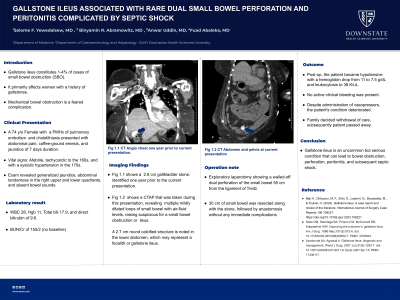
Case Description/Methods: A 74-year-old female with a history of pulmonary embolism and cholelithiasis presented following several days of abdominal pain, coffee-ground emesis, and jaundice. She endorsed loss of appetite but denied fever or chills. She was afebrile, tachycardic to the 160s, and with a SBP in the 170s. A physical exam revealed generalized jaundice, abdominal tenderness in the right upper and lower quadrants, and no audible bowel sounds. Labs showed leukocytosis and mild anemia, total bilirubin of 17.9, direct bilirubin of 9.6, and BUN/Cr of 155/2 (no baseline for comparison). A CT abdomen/pelvis suggested a possible SBO, indicative of gallstone ileus. The patient was given intravenous fluids and started on broad-spectrum antibiotics. An NG tube was inserted for decompression, yielding minimal brownish fluid. An exploratory laparotomy was performed, showing a walled-off dual perforation of the small bowel 50 cm from the ligament of Treitz caused by the obstructing stone. 30 cm of small bowel was resected along with the stone, followed by anastomosis without any immediate complications. Postoperatively, the patient became hypotensive with a hemoglobin drop from 11 to 7.5 g/dL and leukocytosis to 38 K/uL. No active clinical bleeding was present. Despite the administration of pressor, the patient's condition deteriorated. The family decided on the withdrawal of care, and subsequently, the patient passed away.
Discussion: Gallstone ileus is an uncommon but serious condition that can lead to bowel perforation, peritonitis, and subsequent septic shock. Gastroenterologists should familiarize themselves with this clinical entity to recognize and intervene to prevent this life-threatening complication. Further research is needed to determine standardized guidelines on revised indications for prophylactic cholecystectomy to avert bad outcomes.

Disclosures:
Selome F. Yewedalsew, MD1, Binyamin R. Abramowitz, MD2, Anwar Uddin, MD3, Fuad Abaleka, MD4, Tamta Chkhikvadze, MD1. P3510 - Gallstone Ileus-Associated With Rare Dual Small Bowel Perforation and Peritonitis Complicated by Septic Shock, ACG 2024 Annual Scientific Meeting Abstracts. Philadelphia, PA: American College of Gastroenterology.
1SUNY Downstate Health Sciences University, Brooklyn, NY; 2SUNY Downstate Health Sciences University, New York, NY; 3SUNY Downstate Medical Center, Brooklyn, NY; 4Covenant Medical Center, Lubbock, TX
Introduction: Gallstone ileus is a rare condition constituting 1-4% of cases of small bowel obstruction (SBO). It primarily affects women with a history of gallstones. Mechanical bowel obstruction is a feared complication. Patients typically present with abdominal pain, jaundice, nausea, and vomiting. Diagnosis is often confirmed by a CT abdomen, although serial abdominal X-rays may also be useful. We present a case of a patient presenting with SBO and hyperbilirubinemia, without evident biliary pathology on imaging but with a surgical diagnosis of SBO secondary to a gallstone. This case highlights the diagnostic and treatment challenges in such patients.
Case Description/Methods: A 74-year-old female with a history of pulmonary embolism and cholelithiasis presented following several days of abdominal pain, coffee-ground emesis, and jaundice. She endorsed loss of appetite but denied fever or chills. She was afebrile, tachycardic to the 160s, and with a SBP in the 170s. A physical exam revealed generalized jaundice, abdominal tenderness in the right upper and lower quadrants, and no audible bowel sounds. Labs showed leukocytosis and mild anemia, total bilirubin of 17.9, direct bilirubin of 9.6, and BUN/Cr of 155/2 (no baseline for comparison). A CT abdomen/pelvis suggested a possible SBO, indicative of gallstone ileus. The patient was given intravenous fluids and started on broad-spectrum antibiotics. An NG tube was inserted for decompression, yielding minimal brownish fluid. An exploratory laparotomy was performed, showing a walled-off dual perforation of the small bowel 50 cm from the ligament of Treitz caused by the obstructing stone. 30 cm of small bowel was resected along with the stone, followed by anastomosis without any immediate complications. Postoperatively, the patient became hypotensive with a hemoglobin drop from 11 to 7.5 g/dL and leukocytosis to 38 K/uL. No active clinical bleeding was present. Despite the administration of pressor, the patient's condition deteriorated. The family decided on the withdrawal of care, and subsequently, the patient passed away.
Discussion: Gallstone ileus is an uncommon but serious condition that can lead to bowel perforation, peritonitis, and subsequent septic shock. Gastroenterologists should familiarize themselves with this clinical entity to recognize and intervene to prevent this life-threatening complication. Further research is needed to determine standardized guidelines on revised indications for prophylactic cholecystectomy to avert bad outcomes.

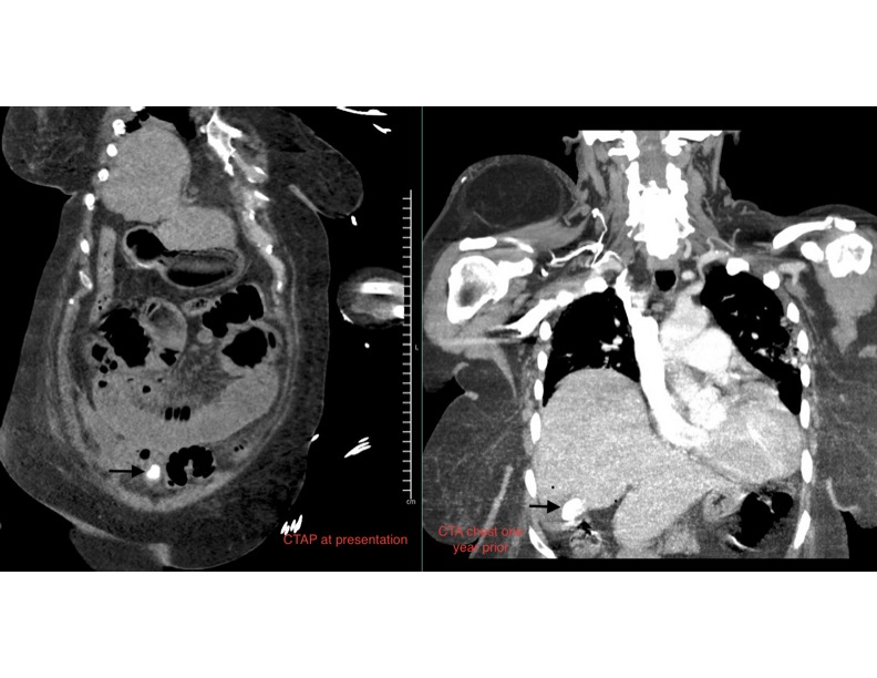
Figure: A prior CT scan showed a calcified gallstone similar to the one now located in the lower pelvis in repeat imaging
Disclosures:
Selome Yewedalsew indicated no relevant financial relationships.
Binyamin Abramowitz indicated no relevant financial relationships.
Anwar Uddin indicated no relevant financial relationships.
Fuad Abaleka indicated no relevant financial relationships.
Tamta Chkhikvadze indicated no relevant financial relationships.
Selome F. Yewedalsew, MD1, Binyamin R. Abramowitz, MD2, Anwar Uddin, MD3, Fuad Abaleka, MD4, Tamta Chkhikvadze, MD1. P3510 - Gallstone Ileus-Associated With Rare Dual Small Bowel Perforation and Peritonitis Complicated by Septic Shock, ACG 2024 Annual Scientific Meeting Abstracts. Philadelphia, PA: American College of Gastroenterology.
