Tuesday Poster Session
Category: Biliary/Pancreas
P3514 - Retained Video Capsule for Anemia Work-Up Diagnoses New Pancreatic Cancer
Tuesday, October 29, 2024
10:30 AM - 4:00 PM ET
Location: Exhibit Hall E

Has Audio

Aria Jalalian, MD
Lankenau Medical Center
Wynnewood, PA
Presenting Author(s)
Aria Jalalian, MD, Stefanie Gallagher, DO, Bryan Stone, DO, Erin Hollis, DO, Malini Mathur, MD
Lankenau Medical Center, Wynnewood, PA
Introduction: Pancreatic cancer often presents as advanced disease, with 47% of patients displaying distant metastasis and 29% local disease at diagnosis. Symptoms typically include abdominal pain, weight loss, and jaundice. Rarely, patients present with asymptomatic metastatic disease. We present a case of a patient undergoing workup for iron deficiency anemia (IDA), who had unremarkable EGD/colonoscopy and subsequently underwent video capsule endoscopy (VCE). The capsule lodged in the small bowel causing obstruction, necessitating a diagnostic laparoscopy which revealed a new diagnosis of primary pancreatic cancer with peritoneal carcinomatosis.
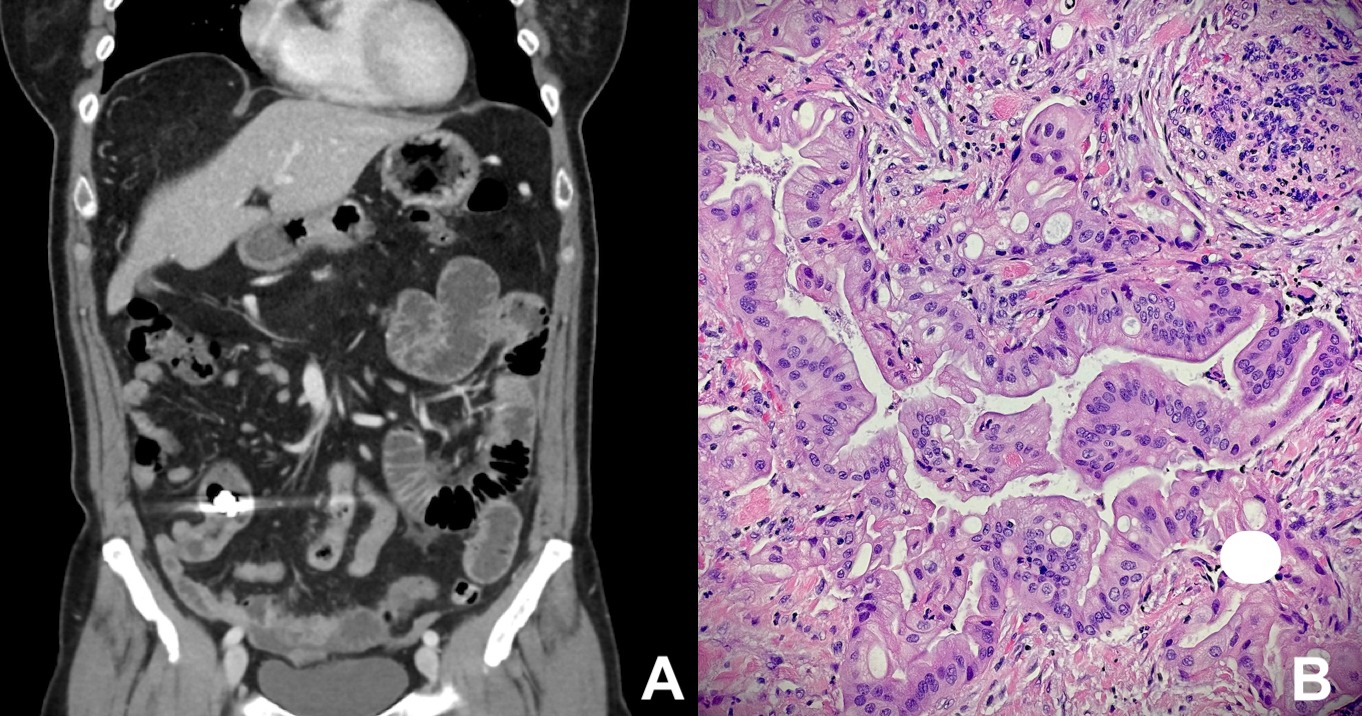
Case Description/Methods: A 62-year-old female with history of polycythemia vera (PCV) presented with days of worsening fatigue and dark stools. Labs showed severe IDA with hemoglobin 5.1 (baseline 14), transferrin saturation 2% and ferritin 1 ng/mL. Her PCV remained stable on hydroxyurea without phlebotomies. Abdominal exam was negative for tenderness, and digital rectal exam showed brown, hemoccult positive stool. CT A/P revealed a pancreatic tail cyst without obvious cause for anemia. She underwent bidirectional endoscopic evaluation to the terminal ileum with no source of IDA identified. She received 3 units of blood and was discharged with outpatient VCE. She returned several days later with worsening periumbilical abdominal pain, nausea and poor appetite. CT A/P revealed small bowel obstruction with transition point proximal to an intraluminal foreign body, the retained capsule. Exploratory laporoscopy revealed extraluminal carcinomatous nodular tissue invading the small bowel wall causing stenosis. Pathology demonstrated adenocarcinoma consistent with pancreaticobiliary origin. CA 19-9 level was 222 U/mL. MRI abdomen showed distal pancreatic body/tail mass abutting the adjacent splenic vessels as well as metastatic peritoneal implants. She was referred to medical and radiation oncology for treatment.
Discussion: This case highlights an unusual presentation of pancreatic cancer, as the patient lacked classical symptoms such as abdominal pain or jaundice. Instead, she underwent standard evaluation for IDA with VCE which lodged at an extraluminal metastatic cancer nodule infiltrating the small bowel, causing obstruction. Our report marks the first instance in literature of initial diagnosis of pancreatic cancer with carcinomatosis through a retained capsule for IDA workup complicated by small bowel obstruction.
Siegel, R. et al., Cancer statistics, 2022, 72(1).

Disclosures:
Aria Jalalian, MD, Stefanie Gallagher, DO, Bryan Stone, DO, Erin Hollis, DO, Malini Mathur, MD. P3514 - Retained Video Capsule for Anemia Work-Up Diagnoses New Pancreatic Cancer, ACG 2024 Annual Scientific Meeting Abstracts. Philadelphia, PA: American College of Gastroenterology.
Lankenau Medical Center, Wynnewood, PA
Introduction: Pancreatic cancer often presents as advanced disease, with 47% of patients displaying distant metastasis and 29% local disease at diagnosis. Symptoms typically include abdominal pain, weight loss, and jaundice. Rarely, patients present with asymptomatic metastatic disease. We present a case of a patient undergoing workup for iron deficiency anemia (IDA), who had unremarkable EGD/colonoscopy and subsequently underwent video capsule endoscopy (VCE). The capsule lodged in the small bowel causing obstruction, necessitating a diagnostic laparoscopy which revealed a new diagnosis of primary pancreatic cancer with peritoneal carcinomatosis.
Case Description/Methods: A 62-year-old female with history of polycythemia vera (PCV) presented with days of worsening fatigue and dark stools. Labs showed severe IDA with hemoglobin 5.1 (baseline 14), transferrin saturation 2% and ferritin 1 ng/mL. Her PCV remained stable on hydroxyurea without phlebotomies. Abdominal exam was negative for tenderness, and digital rectal exam showed brown, hemoccult positive stool. CT A/P revealed a pancreatic tail cyst without obvious cause for anemia. She underwent bidirectional endoscopic evaluation to the terminal ileum with no source of IDA identified. She received 3 units of blood and was discharged with outpatient VCE. She returned several days later with worsening periumbilical abdominal pain, nausea and poor appetite. CT A/P revealed small bowel obstruction with transition point proximal to an intraluminal foreign body, the retained capsule. Exploratory laporoscopy revealed extraluminal carcinomatous nodular tissue invading the small bowel wall causing stenosis. Pathology demonstrated adenocarcinoma consistent with pancreaticobiliary origin. CA 19-9 level was 222 U/mL. MRI abdomen showed distal pancreatic body/tail mass abutting the adjacent splenic vessels as well as metastatic peritoneal implants. She was referred to medical and radiation oncology for treatment.
Discussion: This case highlights an unusual presentation of pancreatic cancer, as the patient lacked classical symptoms such as abdominal pain or jaundice. Instead, she underwent standard evaluation for IDA with VCE which lodged at an extraluminal metastatic cancer nodule infiltrating the small bowel, causing obstruction. Our report marks the first instance in literature of initial diagnosis of pancreatic cancer with carcinomatosis through a retained capsule for IDA workup complicated by small bowel obstruction.
Siegel, R. et al., Cancer statistics, 2022, 72(1).

Figure: Figure 1: (A) CT scan displaying a retained capsule within the ileum with proximal small bowel dilation and thickening of the distal wall. (B) Histology revealing peritoneal fibroadipose tissue infiltration by malignancy confirming the diagnosis of metastatic adenocarcinoma.
Disclosures:
Aria Jalalian indicated no relevant financial relationships.
Stefanie Gallagher indicated no relevant financial relationships.
Bryan Stone indicated no relevant financial relationships.
Erin Hollis indicated no relevant financial relationships.
Malini Mathur indicated no relevant financial relationships.
Aria Jalalian, MD, Stefanie Gallagher, DO, Bryan Stone, DO, Erin Hollis, DO, Malini Mathur, MD. P3514 - Retained Video Capsule for Anemia Work-Up Diagnoses New Pancreatic Cancer, ACG 2024 Annual Scientific Meeting Abstracts. Philadelphia, PA: American College of Gastroenterology.
