Monday Poster Session
Category: Stomach
P3348 - Gastric Duplication Cyst With Stalk in an Elderly Woman: A Case Report of a Rare Discovery
Monday, October 28, 2024
10:30 AM - 4:00 PM ET
Location: Exhibit Hall E


Sharifeh Almasaid, MD
SUNY Upstate Medical University
Syracuse, NY
Presenting Author(s)
Sharifeh Almasaid, MD1, Keshia Suhail, MD2, Arwa Elsamny, MBBCh2, Kelita Singh, MD2
1SUNY Upstate Medical University, Johnson City, TN; 2SUNY Upstate Medical University, Syracuse, NY
Introduction: Gastric duplication cysts are rare congenital anomalies with an incidence between 4-9%. The typical location is along the greater curvature of the stomach with direct attachment to the gastric wall without communication with gastric lumen. They can present with epigastric fullness, weight loss, anemia, dysphagia, abdominal tenderness. However, most tend to be completely asymptomatic and diagnosed incidentally. We present a case of an adult patient with an incidentally discovered gastric duplication cyst with a stalk
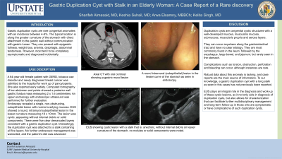
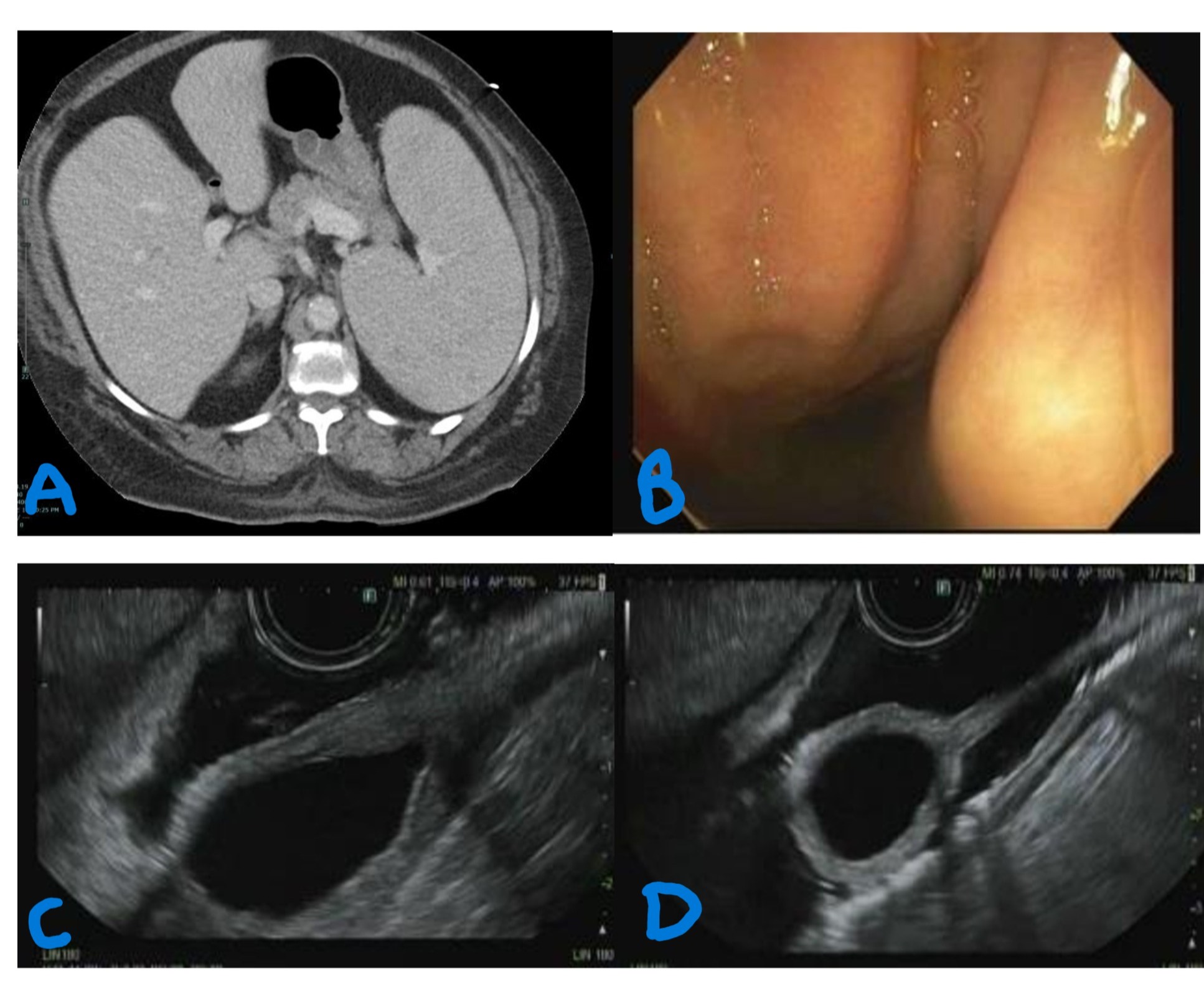
Case Description/Methods: A 64-year-old female patient with GERD, tobacco use disorder and newly diagnosed breast cancer was admitted to the hospital for work up of pancytopenia. She also reported early satiety. Computed tomography of her abdomen and pelvis showed a posterior wall gastric fundus mass measuring 2 x 1.6 centimeters. An upper endoscopy with endoscopic ultrasound (EUS) was performed for further evaluation. Endoscopy revealed a single, non-obstructing, subepithelial lesion with normal overlying mucosa. EUS showed a round, intramural subepithelial lesion in the lesser curvature measuring 18 x 10mm. The lesion was cystic, appearing without internal debris or solid components. There were five clear demarcated layers consistent with a gastric duplication cyst. Interestingly, the duplication cyst was attached to a stalk containing all five layers. No further endoscopic management was warranted, and the patient’s diet was advanced
Discussion: Duplication cysts are congenital cystic structures with a well-developed mucosa, muscularis mucosa, submucosa, muscularis propria and serosa layers. They can occur anywhere along the gastrointestinal tract and have no clear etiology. They are most commonly found in the ileum, followed by the esophagus, large bowel, and jejunum; but rarely seen in the stomach. Complications such as torsion, obstruction, perforation and bleeding can occur, although instances are rare. Robust data about this anomaly is lacking, and case reports are the main source of information. To our knowledge, a gastric duplication cyst with a long stalk as seen in this cases has not previously been reported and the EUS findings are note-worthy as EUS plays an integral role in the diagnosis and work-up of these cystic lesions, as it not only aids in diagnosis of duplication cysts, but also allows for characterization that can facilitate further multidisciplinary management and long-term follow-up in those who are symptomatic or have complications of such duplication cysts

Disclosures:
Sharifeh Almasaid, MD1, Keshia Suhail, MD2, Arwa Elsamny, MBBCh2, Kelita Singh, MD2. P3348 - Gastric Duplication Cyst With Stalk in an Elderly Woman: A Case Report of a Rare Discovery, ACG 2024 Annual Scientific Meeting Abstracts. Philadelphia, PA: American College of Gastroenterology.
1SUNY Upstate Medical University, Johnson City, TN; 2SUNY Upstate Medical University, Syracuse, NY
Introduction: Gastric duplication cysts are rare congenital anomalies with an incidence between 4-9%. The typical location is along the greater curvature of the stomach with direct attachment to the gastric wall without communication with gastric lumen. They can present with epigastric fullness, weight loss, anemia, dysphagia, abdominal tenderness. However, most tend to be completely asymptomatic and diagnosed incidentally. We present a case of an adult patient with an incidentally discovered gastric duplication cyst with a stalk
Case Description/Methods: A 64-year-old female patient with GERD, tobacco use disorder and newly diagnosed breast cancer was admitted to the hospital for work up of pancytopenia. She also reported early satiety. Computed tomography of her abdomen and pelvis showed a posterior wall gastric fundus mass measuring 2 x 1.6 centimeters. An upper endoscopy with endoscopic ultrasound (EUS) was performed for further evaluation. Endoscopy revealed a single, non-obstructing, subepithelial lesion with normal overlying mucosa. EUS showed a round, intramural subepithelial lesion in the lesser curvature measuring 18 x 10mm. The lesion was cystic, appearing without internal debris or solid components. There were five clear demarcated layers consistent with a gastric duplication cyst. Interestingly, the duplication cyst was attached to a stalk containing all five layers. No further endoscopic management was warranted, and the patient’s diet was advanced
Discussion: Duplication cysts are congenital cystic structures with a well-developed mucosa, muscularis mucosa, submucosa, muscularis propria and serosa layers. They can occur anywhere along the gastrointestinal tract and have no clear etiology. They are most commonly found in the ileum, followed by the esophagus, large bowel, and jejunum; but rarely seen in the stomach. Complications such as torsion, obstruction, perforation and bleeding can occur, although instances are rare. Robust data about this anomaly is lacking, and case reports are the main source of information. To our knowledge, a gastric duplication cyst with a long stalk as seen in this cases has not previously been reported and the EUS findings are note-worthy as EUS plays an integral role in the diagnosis and work-up of these cystic lesions, as it not only aids in diagnosis of duplication cysts, but also allows for characterization that can facilitate further multidisciplinary management and long-term follow-up in those who are symptomatic or have complications of such duplication cysts

Figure: Figure A: Axial CT with oral contrast showing a gastric mural lesion
Figure B: A round intramural (subepithelial) lesion in the lesser curve of the stomach.
Figure C,D: The lesion is cystic and anechoic without internal debris, nodules or solid components, seen on lesser curvature.
Figure B: A round intramural (subepithelial) lesion in the lesser curve of the stomach.
Figure C,D: The lesion is cystic and anechoic without internal debris, nodules or solid components, seen on lesser curvature.
Disclosures:
Sharifeh Almasaid indicated no relevant financial relationships.
Keshia Suhail indicated no relevant financial relationships.
Arwa Elsamny indicated no relevant financial relationships.
Kelita Singh indicated no relevant financial relationships.
Sharifeh Almasaid, MD1, Keshia Suhail, MD2, Arwa Elsamny, MBBCh2, Kelita Singh, MD2. P3348 - Gastric Duplication Cyst With Stalk in an Elderly Woman: A Case Report of a Rare Discovery, ACG 2024 Annual Scientific Meeting Abstracts. Philadelphia, PA: American College of Gastroenterology.
