Monday Poster Session
Category: Stomach
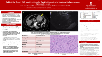
P3351 - Behind the Bleed: EUS Identification of a Gastric Subepithelial Lesion with Spontaneous Hemoperitoneum
Monday, October 28, 2024
10:30 AM - 4:00 PM ET
Location: Exhibit Hall E

Has Audio
- AA
Antonio Alvarez Castaneda, MD
The Ohio State University Wexner Medical Center
Columbus, OH
Presenting Author(s)
Antonio Alvarez Castaneda, MD, Joanna Jiang, MD, Meaghan Costello, MD, Delvise Fogwe, MD, Erica Park, MD
The Ohio State University Wexner Medical Center, Columbus, OH
Introduction: Clinical manifestations of gastric subepithelial lesions are widely variable, ranging from incidentally discovered, asymptomatic lesions to surgical emergencies related to perforation or hemorrhage. Gastric subepithelial lesions have been associated with intraluminal bleeding, but rarely manifest with extraluminal bleeding. In this case, we discuss a unique presentation of a patient with spontaneous hemoperitoneum and the utility of endoscopic ultrasound when there is uncertainty surrounding the source of hemorrhage.
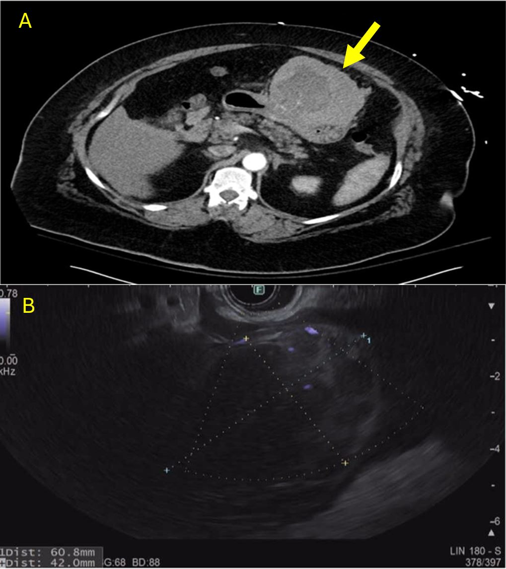
Case Description/Methods: A 57-year-old woman presented with abdominal pain, presyncope and hypotension. Her hemoglobin was decreased to 10 g/dL, and she had significant epigastric pain on exam. After resuscitation, the patient remained hemodynamically stable. Computed tomography angiogram (CTA) of the abdomen and pelvis revealed a large hematoma extending from greater to lesser curvature of the stomach, as well as hemoperitoneum adjacent to liver and spleen without an underlying source (Figure 1A). Due to the unclear etiology of her spontaneous hemoperitoneum and overall clinical stability, the patient underwent endoscopic ultrasound (EUS) for further evaluation. EUS revealed a 6 cm by 4 cm exophytic, highly vascular subepithelial gastric mass in the antrum of the stomach, originating from the muscularis propria. This lesion was contiguous with the surrounding intraperitoneal hematoma, suggestive as the etiology of her hemoperitoneum (Figure 1B). The patient underwent robotic partial gastrectomy with removal of a large, bleeding anterior gastric mass. Patient tolerated the procedure well and had no post operative complications. Histopathological analysis of the mass is pending at the time of abstract submission and will be available at the time of the conference.
Discussion: Initial evaluation and management of spontaneous hemoperitoneum includes resuscitation as needed, followed by identification and management of the causative etiology. In this case, CTA did not identify a clear etiology of spontaneous hemoperitoneum. Endoscopic evaluation with EUS was therefore a crucial diagnostic tool that successfully identified a large subepithelial gastric mass causing this patient’s hemoperitoneum. Management of these lesions includes surgical or endoscopic resection based on the size and pathology of the lesion.

Disclosures:
Antonio Alvarez Castaneda, MD, Joanna Jiang, MD, Meaghan Costello, MD, Delvise Fogwe, MD, Erica Park, MD. P3351 - Behind the Bleed: EUS Identification of a Gastric Subepithelial Lesion with Spontaneous Hemoperitoneum, ACG 2024 Annual Scientific Meeting Abstracts. Philadelphia, PA: American College of Gastroenterology.
The Ohio State University Wexner Medical Center, Columbus, OH
Introduction: Clinical manifestations of gastric subepithelial lesions are widely variable, ranging from incidentally discovered, asymptomatic lesions to surgical emergencies related to perforation or hemorrhage. Gastric subepithelial lesions have been associated with intraluminal bleeding, but rarely manifest with extraluminal bleeding. In this case, we discuss a unique presentation of a patient with spontaneous hemoperitoneum and the utility of endoscopic ultrasound when there is uncertainty surrounding the source of hemorrhage.
Case Description/Methods: A 57-year-old woman presented with abdominal pain, presyncope and hypotension. Her hemoglobin was decreased to 10 g/dL, and she had significant epigastric pain on exam. After resuscitation, the patient remained hemodynamically stable. Computed tomography angiogram (CTA) of the abdomen and pelvis revealed a large hematoma extending from greater to lesser curvature of the stomach, as well as hemoperitoneum adjacent to liver and spleen without an underlying source (Figure 1A). Due to the unclear etiology of her spontaneous hemoperitoneum and overall clinical stability, the patient underwent endoscopic ultrasound (EUS) for further evaluation. EUS revealed a 6 cm by 4 cm exophytic, highly vascular subepithelial gastric mass in the antrum of the stomach, originating from the muscularis propria. This lesion was contiguous with the surrounding intraperitoneal hematoma, suggestive as the etiology of her hemoperitoneum (Figure 1B). The patient underwent robotic partial gastrectomy with removal of a large, bleeding anterior gastric mass. Patient tolerated the procedure well and had no post operative complications. Histopathological analysis of the mass is pending at the time of abstract submission and will be available at the time of the conference.
Discussion: Initial evaluation and management of spontaneous hemoperitoneum includes resuscitation as needed, followed by identification and management of the causative etiology. In this case, CTA did not identify a clear etiology of spontaneous hemoperitoneum. Endoscopic evaluation with EUS was therefore a crucial diagnostic tool that successfully identified a large subepithelial gastric mass causing this patient’s hemoperitoneum. Management of these lesions includes surgical or endoscopic resection based on the size and pathology of the lesion.

Figure: Figure 1: (A) CTA abdomen pelvis showing hematoma (yellow arrow) adjacent to the stomach, causing mass effect posteriorly, (B) EUS image with Doppler demonstrating a vascular, heterogenous, subepithelial mass measuring 60.8 mm x 42.0 mm with surrounding hemoperitoneum.
Disclosures:
Antonio Alvarez Castaneda indicated no relevant financial relationships.
Joanna Jiang indicated no relevant financial relationships.
Meaghan Costello indicated no relevant financial relationships.
Delvise Fogwe indicated no relevant financial relationships.
Erica Park indicated no relevant financial relationships.
Antonio Alvarez Castaneda, MD, Joanna Jiang, MD, Meaghan Costello, MD, Delvise Fogwe, MD, Erica Park, MD. P3351 - Behind the Bleed: EUS Identification of a Gastric Subepithelial Lesion with Spontaneous Hemoperitoneum, ACG 2024 Annual Scientific Meeting Abstracts. Philadelphia, PA: American College of Gastroenterology.
