Monday Poster Session
Category: Stomach
P3366 - Primary Gastric Melanoma Presenting as a Submucosal Lesion
Monday, October 28, 2024
10:30 AM - 4:00 PM ET
Location: Exhibit Hall E

Has Audio

Sareena Ali, DO
Advocate Lutheran General Hospital
Park Ridge, IL
Presenting Author(s)
Sareena Ali, DO1, Robin David, MD2, Nahren Asado, MBChB, MD3, Julio Cabrera, MD1, Asif Lakha, MD1
1Advocate Lutheran General Hospital, Park Ridge, IL; 2Advocate Lutheran General, Chicago, IL; 3Advocate Lutheran General, Park Ridge, IL
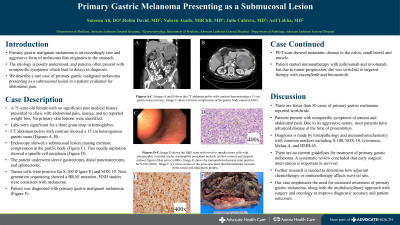
Introduction: Primary gastric malignant melanoma is an exceedingly rare and aggressive form of melanoma that originates in the stomach. The etiology remains poorly understood, and patients often present with nonspecific symptoms which lead to delays in diagnosis. We describe a rare case of primary gastric malignant melanoma presenting as a submucosal lesion in a patient evaluated for abdominal pain.
Case Description/Methods: A 71-year-old female with no significant past medical history presented to clinic with abdominal pain and nausea. She denied weight loss. Her labs showed a three gram drop in hemoglobin from one year prior. Imaging revealed a 15-centimeter heterogenous gastric mass. Endoscopy demonstrated a submucosal lesion causing extrinsic compression at the gastric body. Fine needle aspiration showed a spindle cell neoplasm. The patient underwent sleeve gastrectomy, distal pancreatectomy, and splenectomy. Tumor cells were positive for S-100 and SOX-10. Next generation sequencing showed a BRAF mutation. FISH studies were consistent with melanoma. No primary skin lesions were identified. The patient was diagnosed with primary gastric malignant melanoma. PET scan showed metastatic disease to the colon, small bowel, and muscle. She started immunotherapy with ipilimumab and nivolumab with tumor progression and was switched to targeted therapy with encorafenib and binimetinib.
Discussion: There are fewer than 50 cases of primary gastric melanoma reported worldwide. Patients present with nonspecific symptoms like abdominal pain and nausea. Due to its aggressive nature, most patients have advanced disease at the time of presentation. Diagnosis is made by histopathology and immunohistochemistry with common markers including S-100, SOX-10, tyrosinase, Melan-A, and HMB-45. There are no current guidelines for treatment of primary gastric melanoma. Based on a systematic review, it was concluded that early surgical intervention is important in survival. Further research is needed to determine how adjuvant chemotherapy or immunotherapy affects survival rate. Our case emphasizes the need for increased awareness of primary gastric melanoma, along with the multidisciplinary approach with surgery and oncology to improve diagnostic accuracy and patient outcomes.

Disclosures:
Sareena Ali, DO1, Robin David, MD2, Nahren Asado, MBChB, MD3, Julio Cabrera, MD1, Asif Lakha, MD1. P3366 - Primary Gastric Melanoma Presenting as a Submucosal Lesion, ACG 2024 Annual Scientific Meeting Abstracts. Philadelphia, PA: American College of Gastroenterology.
1Advocate Lutheran General Hospital, Park Ridge, IL; 2Advocate Lutheran General, Chicago, IL; 3Advocate Lutheran General, Park Ridge, IL
Introduction: Primary gastric malignant melanoma is an exceedingly rare and aggressive form of melanoma that originates in the stomach. The etiology remains poorly understood, and patients often present with nonspecific symptoms which lead to delays in diagnosis. We describe a rare case of primary gastric malignant melanoma presenting as a submucosal lesion in a patient evaluated for abdominal pain.
Case Description/Methods: A 71-year-old female with no significant past medical history presented to clinic with abdominal pain and nausea. She denied weight loss. Her labs showed a three gram drop in hemoglobin from one year prior. Imaging revealed a 15-centimeter heterogenous gastric mass. Endoscopy demonstrated a submucosal lesion causing extrinsic compression at the gastric body. Fine needle aspiration showed a spindle cell neoplasm. The patient underwent sleeve gastrectomy, distal pancreatectomy, and splenectomy. Tumor cells were positive for S-100 and SOX-10. Next generation sequencing showed a BRAF mutation. FISH studies were consistent with melanoma. No primary skin lesions were identified. The patient was diagnosed with primary gastric malignant melanoma. PET scan showed metastatic disease to the colon, small bowel, and muscle. She started immunotherapy with ipilimumab and nivolumab with tumor progression and was switched to targeted therapy with encorafenib and binimetinib.
Discussion: There are fewer than 50 cases of primary gastric melanoma reported worldwide. Patients present with nonspecific symptoms like abdominal pain and nausea. Due to its aggressive nature, most patients have advanced disease at the time of presentation. Diagnosis is made by histopathology and immunohistochemistry with common markers including S-100, SOX-10, tyrosinase, Melan-A, and HMB-45. There are no current guidelines for treatment of primary gastric melanoma. Based on a systematic review, it was concluded that early surgical intervention is important in survival. Further research is needed to determine how adjuvant chemotherapy or immunotherapy affects survival rate. Our case emphasizes the need for increased awareness of primary gastric melanoma, along with the multidisciplinary approach with surgery and oncology to improve diagnostic accuracy and patient outcomes.

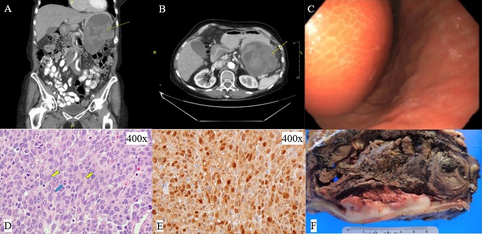
Figure: Figure 1: Image A and B shows CT abdomen pelvis with contrast demonstrating a 15 cm gastric mass (arrows). Image C shows extrinsic compression of the gastric body noted on EGD. Image D shows the H&E stain with ovoid to spindle tumor cells with pleomorphic vesicular nuclei, eosinophilic prominent nucleoli (yellow arrows) and atypical mitoses figures (blue arrow) (400x). Image E shows the immunohistochemical stain positive for S-100 (400x). Image F is a cross-section of the gross specimen that demonstrates invasion of the serosa and muscularis propria.
Disclosures:
Sareena Ali indicated no relevant financial relationships.
Robin David indicated no relevant financial relationships.
Nahren Asado indicated no relevant financial relationships.
Julio Cabrera indicated no relevant financial relationships.
Asif Lakha indicated no relevant financial relationships.
Sareena Ali, DO1, Robin David, MD2, Nahren Asado, MBChB, MD3, Julio Cabrera, MD1, Asif Lakha, MD1. P3366 - Primary Gastric Melanoma Presenting as a Submucosal Lesion, ACG 2024 Annual Scientific Meeting Abstracts. Philadelphia, PA: American College of Gastroenterology.

