Monday Poster Session
Category: Small Intestine
P3271 - Now You "CT" It? A Rare Case of Jejunal Diverticulitis
Monday, October 28, 2024
10:30 AM - 4:00 PM ET
Location: Exhibit Hall E

Has Audio

Bharati Dev, DO
Icahn School of Medicine at Mount Sinai Morningside/West
New York, NY
Presenting Author(s)
Bharati Dev, DO1, Jake DeBroff, MD2, Mako Koseki, MD2, Sonia Mehra, MD3, Subhash Chander, MD2
1Icahn School of Medicine at Mount Sinai Morningside/West, New York, NY; 2Mount Sinai Beth Israel, Icahn School of Medicine at Mount Sinai, New York, NY; 3Mount Sinai Morningside, Icahn School of Medicine at Mount Sinai, New York, NY
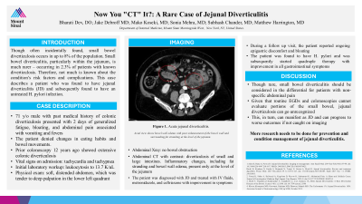
Introduction: Though often incidentally found, small bowel diverticulosis occurs in up to 8% of the population. Small bowel diverticulitis, particularly within the jejunum, is much rarer – occurring in 2.3% of patients with known diverticulosis. Therefore, not much is known about the condition’s risk factors and complications. This case describes a patient with colonic diverticulosis who presented with abdominal pain and was found to have jejunal diverticulitis (JD) in the setting of an untreated H. pylori infection.
Case Description/Methods: A 71-year-old male with a history of colonic diverticulosis presented with 2 days of generalized fatigue, bloating, and abdominal pain associated with vomiting and fevers. Prior to presentation, the patient reported bloating for several months. The patient denied changes in eating habits and bowel movements. Colonoscopy was done 12 years prior which showed extensive colonic diverticulosis.
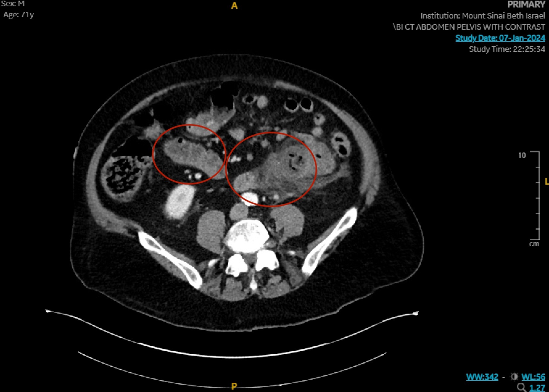
Initial vital signs revealed tachycardia and tachypnea with labs showing leukocytosis to 13.7 K/uL. Exam was notable for a soft, distended abdomen which was tender to palpation in the lower left quadrant. Abdominal x-ray showed no bowel obstruction. CT with contrast of the abdomen revealed diverticulosis of both the small and large intestines. However, inflammatory changes, including fat stranding and bowel wall edema, were only present at the level of the jejunum. The patient was diagnosed with JD and received intravenous fluids, metronidazole, and ceftriaxone, with improvement in abdominal pain.
However, during a follow-up visit, the patient reported ongoing epigastric discomfort and bloating. H. pylori testing was positive, and the patient subsequently started quadruple therapy with improvement in all gastrointestinal symptoms.
Discussion: Though rare, small bowel diverticulitis should be considered in the differential for patients with non-specific abdominal pain. Given that routine endoscopies and colonoscopies cannot evaluate significant portions of the small bowel, jejunal diverticulosis can go unrecognized in patients with extensive colonic diverticulosis.
In turn, inflammation or infection within jejunal diverticula can manifest as JD and clinical outlook could progressively worsen if not caught early. Interestingly in our case, the patient was found to have H. Pylori, which may have been a source for the JD. More research needs to be done for both JD and the impact of H. pylori on small bowel diverticula.

Disclosures:
Bharati Dev, DO1, Jake DeBroff, MD2, Mako Koseki, MD2, Sonia Mehra, MD3, Subhash Chander, MD2. P3271 - Now You "CT" It? A Rare Case of Jejunal Diverticulitis, ACG 2024 Annual Scientific Meeting Abstracts. Philadelphia, PA: American College of Gastroenterology.
1Icahn School of Medicine at Mount Sinai Morningside/West, New York, NY; 2Mount Sinai Beth Israel, Icahn School of Medicine at Mount Sinai, New York, NY; 3Mount Sinai Morningside, Icahn School of Medicine at Mount Sinai, New York, NY
Introduction: Though often incidentally found, small bowel diverticulosis occurs in up to 8% of the population. Small bowel diverticulitis, particularly within the jejunum, is much rarer – occurring in 2.3% of patients with known diverticulosis. Therefore, not much is known about the condition’s risk factors and complications. This case describes a patient with colonic diverticulosis who presented with abdominal pain and was found to have jejunal diverticulitis (JD) in the setting of an untreated H. pylori infection.
Case Description/Methods: A 71-year-old male with a history of colonic diverticulosis presented with 2 days of generalized fatigue, bloating, and abdominal pain associated with vomiting and fevers. Prior to presentation, the patient reported bloating for several months. The patient denied changes in eating habits and bowel movements. Colonoscopy was done 12 years prior which showed extensive colonic diverticulosis.
Initial vital signs revealed tachycardia and tachypnea with labs showing leukocytosis to 13.7 K/uL. Exam was notable for a soft, distended abdomen which was tender to palpation in the lower left quadrant. Abdominal x-ray showed no bowel obstruction. CT with contrast of the abdomen revealed diverticulosis of both the small and large intestines. However, inflammatory changes, including fat stranding and bowel wall edema, were only present at the level of the jejunum. The patient was diagnosed with JD and received intravenous fluids, metronidazole, and ceftriaxone, with improvement in abdominal pain.
However, during a follow-up visit, the patient reported ongoing epigastric discomfort and bloating. H. pylori testing was positive, and the patient subsequently started quadruple therapy with improvement in all gastrointestinal symptoms.
Discussion: Though rare, small bowel diverticulitis should be considered in the differential for patients with non-specific abdominal pain. Given that routine endoscopies and colonoscopies cannot evaluate significant portions of the small bowel, jejunal diverticulosis can go unrecognized in patients with extensive colonic diverticulosis.
In turn, inflammation or infection within jejunal diverticula can manifest as JD and clinical outlook could progressively worsen if not caught early. Interestingly in our case, the patient was found to have H. Pylori, which may have been a source for the JD. More research needs to be done for both JD and the impact of H. pylori on small bowel diverticula.

Figure: Figure 1. Acute jejunal diverticulitis.
Axial view shows bowel wall edema with poor enhancement of the bowel wall and surrounding fat stranding at the level of the jejunum.
Axial view shows bowel wall edema with poor enhancement of the bowel wall and surrounding fat stranding at the level of the jejunum.
Disclosures:
Bharati Dev indicated no relevant financial relationships.
Jake DeBroff indicated no relevant financial relationships.
Mako Koseki indicated no relevant financial relationships.
Sonia Mehra indicated no relevant financial relationships.
Subhash Chander indicated no relevant financial relationships.
Bharati Dev, DO1, Jake DeBroff, MD2, Mako Koseki, MD2, Sonia Mehra, MD3, Subhash Chander, MD2. P3271 - Now You "CT" It? A Rare Case of Jejunal Diverticulitis, ACG 2024 Annual Scientific Meeting Abstracts. Philadelphia, PA: American College of Gastroenterology.
