Monday Poster Session
Category: Small Intestine
P3279 - Rare Roadblock, EUS Leads the Way! Leiomyosarcoma Involving the Duodenum Causing Gastric Outlet Obstruction
Monday, October 28, 2024
10:30 AM - 4:00 PM ET
Location: Exhibit Hall E

Has Audio

Samuel H. Cheong, DO
Banner - University of Arizona
Tucson, AZ
Presenting Author(s)
Samuel H. Cheong, DO1, Brandon Witten, MD2, Rama Mouhaffel, MD1, Diane Rodden, MD2, Alexis Elliott, MD2, Gebran Abboud, MD2
1Banner - University of Arizona, Tucson, AZ; 2University of Arizona College of Medicine, Tucson, AZ
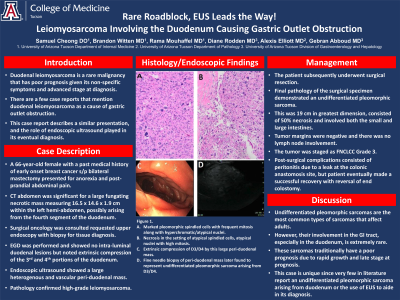
Introduction: Duodenal leiomyosarcoma is a relatively rare malignancy that has poor prognosis given its non-specific symptoms and advanced stage at diagnosis. There are a few case reports that mention duodenal leiomyosarcoma as a cause of gastric outlet obstruction. In this case report, we describe a similar presentation and the role endoscopic ultrasound played in its eventual diagnosis.
Case Description/Methods: A 66 year old female with a past medical history of early onset breast cancer status post bilateral mastectomy presented to the emergency department for anorexia and post-prandial abdominal pain. CT was significant for a large fungating necrotic mass measuring 16.5 x 14.6 x 1.9 cm within the left hemiabdomen, possibly arising from the fourth segment of the duodenum.
Surgical oncology was consulted and requested an upper endoscopy with biopsies for tissue diagnosis. An esophagogastroduodenoscopy (EGD) was performed and showed no intra-luminal duodenal lesions but noted extrinsic compression of the third and fourth portions of the duodenum. Endoscopic ultrasound (EUS) showed a large heterogeneous and vascular peri-duodenal mass. Fine needle biopsies of this mass were obtained using a 22 gauge SharkCore needle. Pathology confirmed a high-grade leiomyosarcoma.
The patient subsequently underwent surgical resection. Final pathology of the surgical specimen determined this was an undifferentiated pleomorphic sarcoma. It was 19cm in greatest dimension, consisted of 50% necrosis, and involved both the small and large intestines. Tumor margins were negative and there was no lymph node involvement. The tumor was staged as FNCLCC Grade 3. Post-surgical complications consisted of peritonitis due to a leak at the colonic anastomosis site, however the patient eventually made a successful recovery with reversal of end colostomy.
Discussion: Undifferentiated pleomorphic sarcomas are the most common types of sarcomas that affect adults. However their involvement in the GI tract, especially the duodenum, is extremely rare. These sarcomas traditionally have a poor prognosis due to rapid growth and late stage at diagnosis. Our finding is unique since very few cases in the literature report an undifferentiated pleomorphic sarcoma arising from duodenum or the use of EUS to aid in its diagnosis.

Disclosures:
Samuel H. Cheong, DO1, Brandon Witten, MD2, Rama Mouhaffel, MD1, Diane Rodden, MD2, Alexis Elliott, MD2, Gebran Abboud, MD2. P3279 - Rare Roadblock, EUS Leads the Way! Leiomyosarcoma Involving the Duodenum Causing Gastric Outlet Obstruction, ACG 2024 Annual Scientific Meeting Abstracts. Philadelphia, PA: American College of Gastroenterology.
1Banner - University of Arizona, Tucson, AZ; 2University of Arizona College of Medicine, Tucson, AZ
Introduction: Duodenal leiomyosarcoma is a relatively rare malignancy that has poor prognosis given its non-specific symptoms and advanced stage at diagnosis. There are a few case reports that mention duodenal leiomyosarcoma as a cause of gastric outlet obstruction. In this case report, we describe a similar presentation and the role endoscopic ultrasound played in its eventual diagnosis.
Case Description/Methods: A 66 year old female with a past medical history of early onset breast cancer status post bilateral mastectomy presented to the emergency department for anorexia and post-prandial abdominal pain. CT was significant for a large fungating necrotic mass measuring 16.5 x 14.6 x 1.9 cm within the left hemiabdomen, possibly arising from the fourth segment of the duodenum.
Surgical oncology was consulted and requested an upper endoscopy with biopsies for tissue diagnosis. An esophagogastroduodenoscopy (EGD) was performed and showed no intra-luminal duodenal lesions but noted extrinsic compression of the third and fourth portions of the duodenum. Endoscopic ultrasound (EUS) showed a large heterogeneous and vascular peri-duodenal mass. Fine needle biopsies of this mass were obtained using a 22 gauge SharkCore needle. Pathology confirmed a high-grade leiomyosarcoma.
The patient subsequently underwent surgical resection. Final pathology of the surgical specimen determined this was an undifferentiated pleomorphic sarcoma. It was 19cm in greatest dimension, consisted of 50% necrosis, and involved both the small and large intestines. Tumor margins were negative and there was no lymph node involvement. The tumor was staged as FNCLCC Grade 3. Post-surgical complications consisted of peritonitis due to a leak at the colonic anastomosis site, however the patient eventually made a successful recovery with reversal of end colostomy.
Discussion: Undifferentiated pleomorphic sarcomas are the most common types of sarcomas that affect adults. However their involvement in the GI tract, especially the duodenum, is extremely rare. These sarcomas traditionally have a poor prognosis due to rapid growth and late stage at diagnosis. Our finding is unique since very few cases in the literature report an undifferentiated pleomorphic sarcoma arising from duodenum or the use of EUS to aid in its diagnosis.

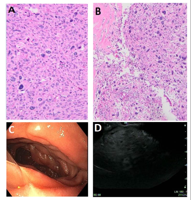
Figure: A: Marked pleomorphic spindled cells with frequent mitosis along with hyperchromatic/atypical nuclei.
B: Necrosis in the setting of atypical spindled cells, atypical nuclei with high mitosis.
C: Extrinsic Compression of D3/D4 by this large peri-duodenal mass.
D: Fine needle biopsy of periduodenal mass later found to represent undifferentiated pleomorphic sarcoma arising from D3/D4.
B: Necrosis in the setting of atypical spindled cells, atypical nuclei with high mitosis.
C: Extrinsic Compression of D3/D4 by this large peri-duodenal mass.
D: Fine needle biopsy of periduodenal mass later found to represent undifferentiated pleomorphic sarcoma arising from D3/D4.
Disclosures:
Samuel Cheong indicated no relevant financial relationships.
Brandon Witten indicated no relevant financial relationships.
Rama Mouhaffel indicated no relevant financial relationships.
Diane Rodden indicated no relevant financial relationships.
Alexis Elliott indicated no relevant financial relationships.
Gebran Abboud indicated no relevant financial relationships.
Samuel H. Cheong, DO1, Brandon Witten, MD2, Rama Mouhaffel, MD1, Diane Rodden, MD2, Alexis Elliott, MD2, Gebran Abboud, MD2. P3279 - Rare Roadblock, EUS Leads the Way! Leiomyosarcoma Involving the Duodenum Causing Gastric Outlet Obstruction, ACG 2024 Annual Scientific Meeting Abstracts. Philadelphia, PA: American College of Gastroenterology.
