Monday Poster Session
Category: Small Intestine
P3223 - MAC Attack: Another Cause of Villous Bunting in a Young Adult with HIV
Monday, October 28, 2024
10:30 AM - 4:00 PM ET
Location: Exhibit Hall E

Has Audio

Keerthi Thallapureddy, MD
University of Texas Health San Antonio
San Antonio, TX
Presenting Author(s)
Keerthi Thallapureddy, MD1, Saatchi Kuwelker, MD2, Carl Kay, MD3, Nagasri Shankar, MD1, Kenneth Hughes, MD1, Randy Wright, MD1
1University of Texas Health San Antonio, San Antonio, TX; 2University of Texas Health Science Center, San Antonio, TX; 3Carl R. Darnall Army Medical Center, Georgetown, TX
Introduction: Mycobacterium avium complex (MAC) is a group of infections caused by nontuberculous mycobacterial species: M avium and M intracellulare. We present a case of a young man with HIV who was found to have a symptomatic MAC as an AIDS-defining condition.
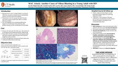
Case Description/Methods: A 26-year-old male with HIV (CD4 count < 40 cells/mm3) on bictegravir/emtricitabine/tenofovir alafenamide presented with 6 months of progressive postprandial left lower quadrant abdominal pain, anorexia, 30-pound weight loss, and iron deficiency anemia. CT imaging revealed hepatosplenomegaly with profound intra-abdominal lymphadenopathy. He underwent bidirectional endoscopy to evaluate iron deficiency anemia. Upper endoscopy found diffuse moderately erythematous, edematous, and friable mucosa in the second portion of the duodenum, that was biopsied (Figure 1). He also underwent ultrasound-guided lymph node biopsy. Duodenal biopsy (Figure 2) revealed small bowel villous blunting. After further staining, both lymph node biopsy and duodenal biopsy revealed concordant evidence of Mycobacterium avium-intracellulare complex infection. His antiviral therapy was changed to dolutegravir and emtricitabine/tenofovir, and he was started on a one-year treatment regimen of rifampin, azithromycin, and ethambutol.
Discussion: Mycobacterium avium complex (MAC) infections are commonly seen in HIV patients with CD4 counts less than 50 cells/mm3. When disseminated, MAC is an AIDS-defining condition. Patients typically present with nonspecific symptoms and often localize to the GI tract including the stomach and duodenum. Endoscopic findings include mucosal edema, friability, erosions/ulcerations, and lymphangiectasis. Histologic findings include patchy diffuse histiocytic infiltrates with lymphoplasmacytic infiltrate and villi broadening in the lamina propria, as seen in this patient. AFB stains are critical in all biopsy specimens from HIV/AIDS to help aid with diagnosis. This case emphasizes the broad differential diagnosis for small bowel villous blunting. Most notably, this case highlights gastrointestinal MAC as a complication of HIV in a young adult presenting with nonspecific GI symptoms.

Disclosures:
Keerthi Thallapureddy, MD1, Saatchi Kuwelker, MD2, Carl Kay, MD3, Nagasri Shankar, MD1, Kenneth Hughes, MD1, Randy Wright, MD1. P3223 - MAC Attack: Another Cause of Villous Bunting in a Young Adult with HIV, ACG 2024 Annual Scientific Meeting Abstracts. Philadelphia, PA: American College of Gastroenterology.
1University of Texas Health San Antonio, San Antonio, TX; 2University of Texas Health Science Center, San Antonio, TX; 3Carl R. Darnall Army Medical Center, Georgetown, TX
Introduction: Mycobacterium avium complex (MAC) is a group of infections caused by nontuberculous mycobacterial species: M avium and M intracellulare. We present a case of a young man with HIV who was found to have a symptomatic MAC as an AIDS-defining condition.
Case Description/Methods: A 26-year-old male with HIV (CD4 count < 40 cells/mm3) on bictegravir/emtricitabine/tenofovir alafenamide presented with 6 months of progressive postprandial left lower quadrant abdominal pain, anorexia, 30-pound weight loss, and iron deficiency anemia. CT imaging revealed hepatosplenomegaly with profound intra-abdominal lymphadenopathy. He underwent bidirectional endoscopy to evaluate iron deficiency anemia. Upper endoscopy found diffuse moderately erythematous, edematous, and friable mucosa in the second portion of the duodenum, that was biopsied (Figure 1). He also underwent ultrasound-guided lymph node biopsy. Duodenal biopsy (Figure 2) revealed small bowel villous blunting. After further staining, both lymph node biopsy and duodenal biopsy revealed concordant evidence of Mycobacterium avium-intracellulare complex infection. His antiviral therapy was changed to dolutegravir and emtricitabine/tenofovir, and he was started on a one-year treatment regimen of rifampin, azithromycin, and ethambutol.
Discussion: Mycobacterium avium complex (MAC) infections are commonly seen in HIV patients with CD4 counts less than 50 cells/mm3. When disseminated, MAC is an AIDS-defining condition. Patients typically present with nonspecific symptoms and often localize to the GI tract including the stomach and duodenum. Endoscopic findings include mucosal edema, friability, erosions/ulcerations, and lymphangiectasis. Histologic findings include patchy diffuse histiocytic infiltrates with lymphoplasmacytic infiltrate and villi broadening in the lamina propria, as seen in this patient. AFB stains are critical in all biopsy specimens from HIV/AIDS to help aid with diagnosis. This case emphasizes the broad differential diagnosis for small bowel villous blunting. Most notably, this case highlights gastrointestinal MAC as a complication of HIV in a young adult presenting with nonspecific GI symptoms.

Figure: Figure 1: Upper endoscopy findings of mucosal edema, friability (1A&1B), erosions and ulcerations (1A), and lymphangiectasia (1A&1B).
Figure 2A&B: Hematoxylin & Eosin slides (40X and 400x) duodenal mucosa with villous blunting and lamina propria with abundant histiocytic infiltrate.
Figures 2C&D: Ziehl-Neelsen stain (40X and 400X) lamina propria histocyte infiltrate with positive staining for innumerable acid-fast bacilli within macrophages consistent with MAI complex.
Disclosures:
Keerthi Thallapureddy indicated no relevant financial relationships.
Saatchi Kuwelker indicated no relevant financial relationships.
Carl Kay indicated no relevant financial relationships.
Nagasri Shankar indicated no relevant financial relationships.
Kenneth Hughes indicated no relevant financial relationships.
Randy Wright indicated no relevant financial relationships.
Keerthi Thallapureddy, MD1, Saatchi Kuwelker, MD2, Carl Kay, MD3, Nagasri Shankar, MD1, Kenneth Hughes, MD1, Randy Wright, MD1. P3223 - MAC Attack: Another Cause of Villous Bunting in a Young Adult with HIV, ACG 2024 Annual Scientific Meeting Abstracts. Philadelphia, PA: American College of Gastroenterology.
