Monday Poster Session
Category: Small Intestine
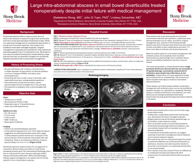
P3224 - Large Intra-Abdominal Abscess in Small Bowel Diverticulitis Treated Nonoperatively Despite Initial Failure With Medical Management
Monday, October 28, 2024
10:30 AM - 4:00 PM ET
Location: Exhibit Hall E

Has Audio

Madeleine Z. Wong, MD
Stony Brook University Hospital
Central Islip, NY
Presenting Author(s)
Madeleine Z. Wong, MD1, John G. Yuen, PhD2, Lindsey Schachter, MD3
1Stony Brook University Hospital, Central Islip, NY; 2Renaissance School of Medicine at Stony Brook University, Stony Brook, NY; 3Stony Brook University Hospital, Stony Brook, NY
Introduction: Small bowel diverticulitis is a less common form of diverticular disease compared to large bowel diverticulitis, and has no gold standard of treatment to date. While non-operative management of complicated diverticulitis in the jejunum has been reported, most cases in the duodenum have been managed surgically. Surgical management is typically recommended for intra-abdominal abscesses greater than 3-5 centimeters (cm). We present a rare case of duodenal diverticulitis complicated by a large abscess that was successfully managed non-operatively despite initial failure with conservative management.
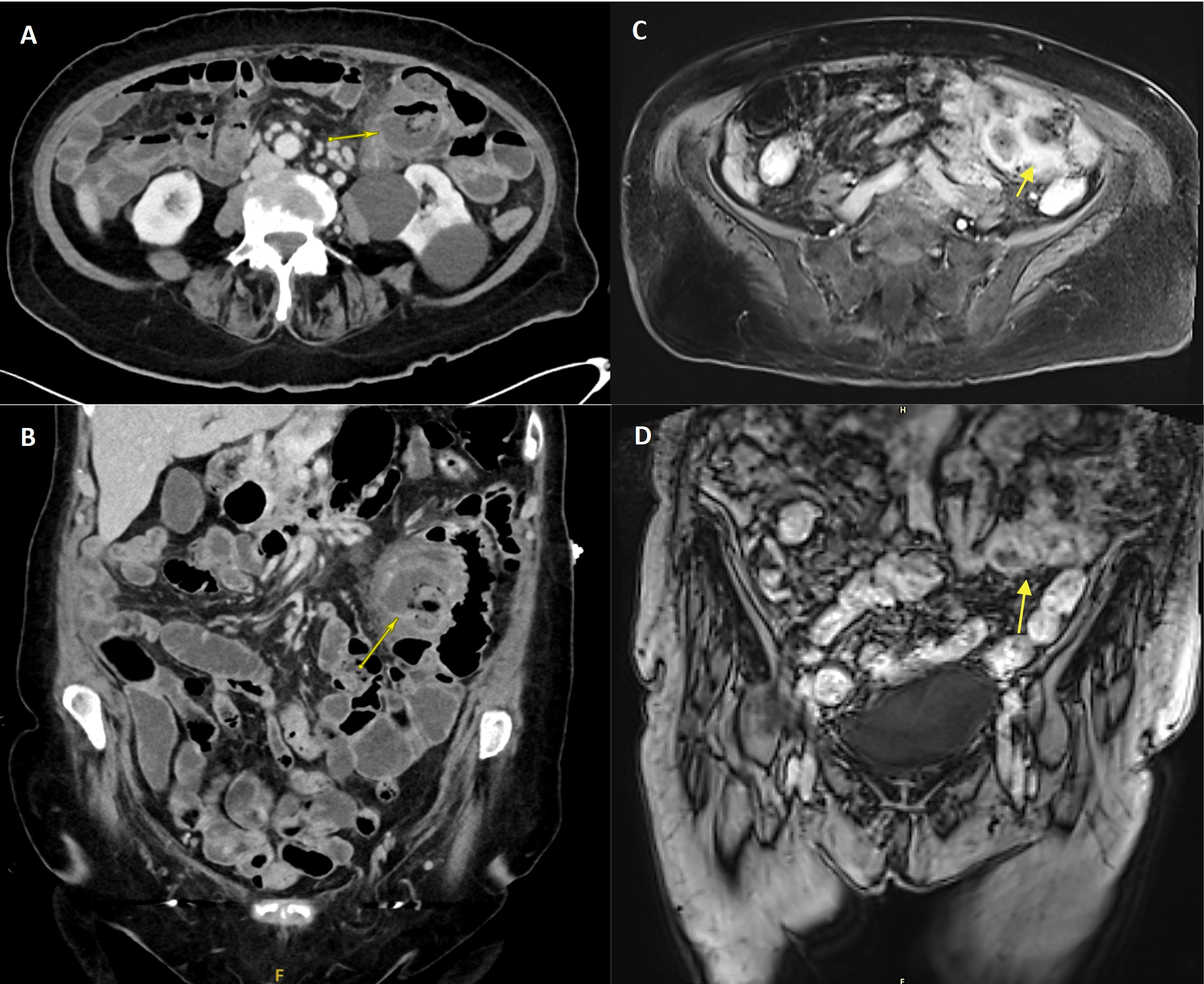
Case Description/Methods: An 80-year-old female was admitted to the hospital for treatment of complicated duodenal diverticulitis. Computed tomography (CT) scan of the abdomen was significant for a 5.5 x 5.0 x 4.1 cm enhancing collection in the duodenum (Image A & B). The patient was initiated on broad-spectrum intravenous (IV) antibiotics. The abscess was anatomically inaccessible for CT-guided drainage. The patient and her family deferred surgery due to her advanced age. CT imaging five days after IV antibiotics showed evidence of a shrinking abscess and she was discharged on oral antibiotics with plans for close follow-up. However, the patient returned to the hospital one week later with worsening symptoms, and CT imaging revealed the abscess collection measuring 3 x 2.3 x 2.5 cm, with a new lateral collection measuring 3.1 x 3.5 x 3.2 cm, either as two adjacent or a single bilobed abscess (Image C & D). The patient continued to defer surgery and was started on three weeks of IV antibiotics with complete resolution of her abscesses.
Discussion: Most patients with acute diverticulitis who fail initial conservative treatment with antibiotics, particularly when abscess drainage is not an option, proceed to surgical management (e.g. resection of the diseased bowel). This case presents a unique situation where a large duodenal diverticular abscess that was not amenable to CT-guided drainage, was successfully resolved with IV antibiotics despite the initial failure of oral antibiotics. Future studies can further assess whether conservative management with antibiotics alone can be risk-stratified by anatomic location (i.e., jejunoileal vs. duodenal). Similarly, studies on whether parenteral or oral antibiotics are more efficacious at treating complicated small bowel diverticulitis may help guide discharge planning for patients in the future.

Disclosures:
Madeleine Z. Wong, MD1, John G. Yuen, PhD2, Lindsey Schachter, MD3. P3224 - Large Intra-Abdominal Abscess in Small Bowel Diverticulitis Treated Nonoperatively Despite Initial Failure With Medical Management, ACG 2024 Annual Scientific Meeting Abstracts. Philadelphia, PA: American College of Gastroenterology.
1Stony Brook University Hospital, Central Islip, NY; 2Renaissance School of Medicine at Stony Brook University, Stony Brook, NY; 3Stony Brook University Hospital, Stony Brook, NY
Introduction: Small bowel diverticulitis is a less common form of diverticular disease compared to large bowel diverticulitis, and has no gold standard of treatment to date. While non-operative management of complicated diverticulitis in the jejunum has been reported, most cases in the duodenum have been managed surgically. Surgical management is typically recommended for intra-abdominal abscesses greater than 3-5 centimeters (cm). We present a rare case of duodenal diverticulitis complicated by a large abscess that was successfully managed non-operatively despite initial failure with conservative management.
Case Description/Methods: An 80-year-old female was admitted to the hospital for treatment of complicated duodenal diverticulitis. Computed tomography (CT) scan of the abdomen was significant for a 5.5 x 5.0 x 4.1 cm enhancing collection in the duodenum (Image A & B). The patient was initiated on broad-spectrum intravenous (IV) antibiotics. The abscess was anatomically inaccessible for CT-guided drainage. The patient and her family deferred surgery due to her advanced age. CT imaging five days after IV antibiotics showed evidence of a shrinking abscess and she was discharged on oral antibiotics with plans for close follow-up. However, the patient returned to the hospital one week later with worsening symptoms, and CT imaging revealed the abscess collection measuring 3 x 2.3 x 2.5 cm, with a new lateral collection measuring 3.1 x 3.5 x 3.2 cm, either as two adjacent or a single bilobed abscess (Image C & D). The patient continued to defer surgery and was started on three weeks of IV antibiotics with complete resolution of her abscesses.
Discussion: Most patients with acute diverticulitis who fail initial conservative treatment with antibiotics, particularly when abscess drainage is not an option, proceed to surgical management (e.g. resection of the diseased bowel). This case presents a unique situation where a large duodenal diverticular abscess that was not amenable to CT-guided drainage, was successfully resolved with IV antibiotics despite the initial failure of oral antibiotics. Future studies can further assess whether conservative management with antibiotics alone can be risk-stratified by anatomic location (i.e., jejunoileal vs. duodenal). Similarly, studies on whether parenteral or oral antibiotics are more efficacious at treating complicated small bowel diverticulitis may help guide discharge planning for patients in the future.

Figure: Contrasted computed tomography of the abdomen showing large, multiloculated duodenal diverticulum arising from the second portion of the duodenum with enhancing fluid collection measuring approximately 5.5 x 4.1 x 5.0 cm (yellow arrow) in transverse view (image A) and coronal view (image B). MRI with contrast or the abdomen showing a medial collection measured 3 x 2.3 x 2.5 cm, and the lateral collection measured 3.1 x 3.5 x 3.2 cm, either as 2 adjacent versus a single bilobed abscess (yellow arrow), in in transverse view (image C) and coronal view (image D)
Disclosures:
Madeleine Wong indicated no relevant financial relationships.
John Yuen indicated no relevant financial relationships.
Lindsey Schachter indicated no relevant financial relationships.
Madeleine Z. Wong, MD1, John G. Yuen, PhD2, Lindsey Schachter, MD3. P3224 - Large Intra-Abdominal Abscess in Small Bowel Diverticulitis Treated Nonoperatively Despite Initial Failure With Medical Management, ACG 2024 Annual Scientific Meeting Abstracts. Philadelphia, PA: American College of Gastroenterology.
