Monday Poster Session
Category: Liver
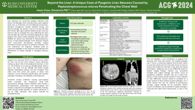
P3022 - Beyond the Liver: A Unique Case of Pyogenic Liver Abscess Caused by P. Micros Penetrating Into the Chest Wall
Monday, October 28, 2024
10:30 AM - 4:00 PM ET
Location: Exhibit Hall E

Has Audio

Isaac Giovannie, MD
Brooklyn Hospital Center
Chicago, IL
Presenting Author(s)
Giovannie Isaac-Coss, MD1, Alex Chow, MD2, Vikash Kumar, MD3, Camelia Ciobanu, MD1, Madhavi Reddy, MD, FACG1, Mohammad Nawaz, MD1
1Brooklyn Hospital Center, Brooklyn, NY; 2SUNY Downstate Health Sciences University, Brooklyn, NY; 3Creighton University School of Medicine, Brooklyn, NY
Introduction: Liver abscesses are uncommon pyogenic infections with highly variable microbiology. Management usually involves the combination of antibiotics and abscess drainage. Here we present a case of a 37 year old male with chronic right upper quadrant abdominal pain with new right chest wall mass and liver mass mistakenly considered to be malignancy and found to have a large pyogenic liver abscess invading into the chest wall. This case also features a rarely isolated pathogen in liver abscess, Peptostreptococcus micros. Additionally, our patient did not respond to traditional percutaneous drainage and required surgical management.
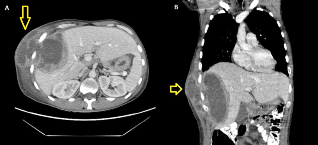
Case Description/Methods: A 37-year-old male that was referred due to an enlarging liver mass infiltrating the chest wall and right-side chest ribs along with severe RUQ pain. On presentations, he was afebrile. Physical exam remarkable for erythematous circular protrusion on right lower chest wall and mild abdominal tenderness. Labs were significant for WBC 16. Originally Abx were not started but the patient became febrile with Tmax 102.8 F and WBC uptrended to 21.9. Antibiotic therapy with vancomycin, cefepime, and metronidazole was started for broad-spectrum coverage. Imaging showed a large collection with extension to the right lateral chest wall (Image 1). Malignancy evaluation w/ AFP, CA 19-9, CEA, all WNL. Percutaneous drainage was conducted without success. Consequently, he underwent surgical drainage. Antibiotics ceased post-surgery due to rapid clinical improvement. Fluid culture analysis subsequently finalized to P. micros.
Discussion: This case highlights a rare complication of pyogenic liver abscesses with chest wall invasion. The isolated pathogen was P micra, a commensal organism that has been shown to cause liver abscess but has never been shown to cause an abscess that invades the chest wall. In fact, given this unique presentation, malignancy was on the differential based on original imaging findings. Infection was successfully managed with surgical drainage and shorter course of appropriate antibiotics. Providers that see a similar liver abscess in their practice should consider P micra infection on their differential. Cultures should be incubated for a longer duration to help cultivate causative pathogens. For a similar abscess that may not respond to percutaneous drainage, surgical drainage can be considered. Shorter courses of antibiotics may be justified if surgical drainage is successful with rapid clinical improvement, as seen in this case.

Note: The table for this abstract can be viewed in the ePoster Gallery section of the ACG 2024 ePoster Site or in The American Journal of Gastroenterology's abstract supplement issue, both of which will be available starting October 27, 2024.
Disclosures:
Giovannie Isaac-Coss, MD1, Alex Chow, MD2, Vikash Kumar, MD3, Camelia Ciobanu, MD1, Madhavi Reddy, MD, FACG1, Mohammad Nawaz, MD1. P3022 - Beyond the Liver: A Unique Case of Pyogenic Liver Abscess Caused by P. Micros Penetrating Into the Chest Wall, ACG 2024 Annual Scientific Meeting Abstracts. Philadelphia, PA: American College of Gastroenterology.
1Brooklyn Hospital Center, Brooklyn, NY; 2SUNY Downstate Health Sciences University, Brooklyn, NY; 3Creighton University School of Medicine, Brooklyn, NY
Introduction: Liver abscesses are uncommon pyogenic infections with highly variable microbiology. Management usually involves the combination of antibiotics and abscess drainage. Here we present a case of a 37 year old male with chronic right upper quadrant abdominal pain with new right chest wall mass and liver mass mistakenly considered to be malignancy and found to have a large pyogenic liver abscess invading into the chest wall. This case also features a rarely isolated pathogen in liver abscess, Peptostreptococcus micros. Additionally, our patient did not respond to traditional percutaneous drainage and required surgical management.
Case Description/Methods: A 37-year-old male that was referred due to an enlarging liver mass infiltrating the chest wall and right-side chest ribs along with severe RUQ pain. On presentations, he was afebrile. Physical exam remarkable for erythematous circular protrusion on right lower chest wall and mild abdominal tenderness. Labs were significant for WBC 16. Originally Abx were not started but the patient became febrile with Tmax 102.8 F and WBC uptrended to 21.9. Antibiotic therapy with vancomycin, cefepime, and metronidazole was started for broad-spectrum coverage. Imaging showed a large collection with extension to the right lateral chest wall (Image 1). Malignancy evaluation w/ AFP, CA 19-9, CEA, all WNL. Percutaneous drainage was conducted without success. Consequently, he underwent surgical drainage. Antibiotics ceased post-surgery due to rapid clinical improvement. Fluid culture analysis subsequently finalized to P. micros.
Discussion: This case highlights a rare complication of pyogenic liver abscesses with chest wall invasion. The isolated pathogen was P micra, a commensal organism that has been shown to cause liver abscess but has never been shown to cause an abscess that invades the chest wall. In fact, given this unique presentation, malignancy was on the differential based on original imaging findings. Infection was successfully managed with surgical drainage and shorter course of appropriate antibiotics. Providers that see a similar liver abscess in their practice should consider P micra infection on their differential. Cultures should be incubated for a longer duration to help cultivate causative pathogens. For a similar abscess that may not respond to percutaneous drainage, surgical drainage can be considered. Shorter courses of antibiotics may be justified if surgical drainage is successful with rapid clinical improvement, as seen in this case.

Figure: CT chest/abdomen/pelvis with IV contrast (sagittal and coronal views) showing a large 10 x 7 x 15 cm hypodense collection with multiple enhancing septa with enhancing capsule. There is mild surrounding hyperenhancement of the liver parenchyma suggesting inflammation. Extension of complex collection abscesses to the right lateral chest wall through the right 7th through 9th intercostal rib spaces.
Note: The table for this abstract can be viewed in the ePoster Gallery section of the ACG 2024 ePoster Site or in The American Journal of Gastroenterology's abstract supplement issue, both of which will be available starting October 27, 2024.
Disclosures:
Giovannie Isaac-Coss indicated no relevant financial relationships.
Alex Chow indicated no relevant financial relationships.
Vikash Kumar indicated no relevant financial relationships.
Camelia Ciobanu indicated no relevant financial relationships.
Madhavi Reddy indicated no relevant financial relationships.
Mohammad Nawaz indicated no relevant financial relationships.
Giovannie Isaac-Coss, MD1, Alex Chow, MD2, Vikash Kumar, MD3, Camelia Ciobanu, MD1, Madhavi Reddy, MD, FACG1, Mohammad Nawaz, MD1. P3022 - Beyond the Liver: A Unique Case of Pyogenic Liver Abscess Caused by P. Micros Penetrating Into the Chest Wall, ACG 2024 Annual Scientific Meeting Abstracts. Philadelphia, PA: American College of Gastroenterology.
