Monday Poster Session
Category: Liver
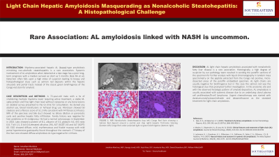
P3026 - Light Chain Hepatic Amyloidosis Masquerading as Nonalcoholic Steatohepatitis: A Histopathological Challenge
Monday, October 28, 2024
10:30 AM - 4:00 PM ET
Location: Exhibit Hall E

Has Audio

Jonathan C. Martinez, MD
St. Francis Hospital
Fortson, GA
Presenting Author(s)
Daniel Schoenborn, DO1, Humberto Rios, MD2, Jonathan C. Martinez, MD3, William F. Willett, MD4, Harold G. Jarrell, MD2
1Southeastern Gastroenterology, Fortson, GA; 2St. Francis Hospital, Columbus, GA; 3St. Francis Hospital, Fortson, GA; 4St. Francis Hospital/ Emory Healthcare, Columbus, GA
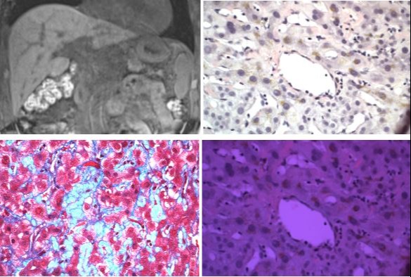
Introduction: Myeloma-associated hepatic AL (kappa)-type amyloidosis mimicking non-alcoholic steatohepatitis is a rare association. Systemic involvement of AL amyloidosis when detected at a late stage has a poor long-term prognosis with a median survival as short as 5 months [Kyle RA et al]. Detection often falls upon a high index of suspicion leading to biopsy and histopathological clues such as salmon red deposits within the hepatic sinusoids and portal tracts instead of the classic green birefringence of the Congo red stain for amyloid.
Case Description/Methods: A 72-year-old male with a hx of smoldering multiple myeloma never requiring active treatment, a stable M-spike protein and free light chain level without cytopenia or any bone lesions on skeletal survey presented to the GI clinic for consultation. He denied any alcohol use, blood transfusions or IV/nasal drug use. FibroScan showed S0 steatosis and a fibrosis score of 51.2 kPa consistent with F4 fibrosis (cirrhosis). MRI of the pancreas and bile duct showed no biliary dilation or pancreatic cysts and positive hepatic fatty infiltration. Family history was negative for liver problems or GI malignancy. He had a normal colonoscopy in September in 2023. Labs showed a protein of 8.1, albumin of 3.5, globulin 4.6, A/G ratio 0.8, T bili 1.5, D bili 0.6,elevated alk-phos 294, AST (SGOT) 65 and ALT (SGPT) 60. EGD showed diffuse, mild, erythematous and friable mucosa with mild portal hypertensive gastropathy found throughout the stomach. CT biopsy of the liver core showed diffuse amyloidosis AL type negative for cirrhosis.
Discussion: AL light chain hepatic amyloidosis associated with nonalcoholic fatty liver disease is a rare association. Histologically, a high degree of suspicion by our pathology team resulted in sending the paraffin sections of the specimen for further analysis with liquid chromatography in tandem mass spectrometry on the peptide extracted from the Congo red positive, micro-dissected areas of the paraffin embedded specimen. AL light chains are usually negative for birefringence but in this case the salmon red was the histological clue that prompted further investigation. In this anatomic site and with the observed histologic pattern of amyloid deposition, AL amyloidosis is usually associated with systemic disease due to an underlying clonal plasma cell proliferation/B-cell lymphoma. Urgent chemotherapy was started with daratumumab/cytoxan/velcade and dexamethasone as the standard treatment for light chain amyloidosis.

Disclosures:
Daniel Schoenborn, DO1, Humberto Rios, MD2, Jonathan C. Martinez, MD3, William F. Willett, MD4, Harold G. Jarrell, MD2. P3026 - Light Chain Hepatic Amyloidosis Masquerading as Nonalcoholic Steatohepatitis: A Histopathological Challenge, ACG 2024 Annual Scientific Meeting Abstracts. Philadelphia, PA: American College of Gastroenterology.
1Southeastern Gastroenterology, Fortson, GA; 2St. Francis Hospital, Columbus, GA; 3St. Francis Hospital, Fortson, GA; 4St. Francis Hospital/ Emory Healthcare, Columbus, GA
Introduction: Myeloma-associated hepatic AL (kappa)-type amyloidosis mimicking non-alcoholic steatohepatitis is a rare association. Systemic involvement of AL amyloidosis when detected at a late stage has a poor long-term prognosis with a median survival as short as 5 months [Kyle RA et al]. Detection often falls upon a high index of suspicion leading to biopsy and histopathological clues such as salmon red deposits within the hepatic sinusoids and portal tracts instead of the classic green birefringence of the Congo red stain for amyloid.
Case Description/Methods: A 72-year-old male with a hx of smoldering multiple myeloma never requiring active treatment, a stable M-spike protein and free light chain level without cytopenia or any bone lesions on skeletal survey presented to the GI clinic for consultation. He denied any alcohol use, blood transfusions or IV/nasal drug use. FibroScan showed S0 steatosis and a fibrosis score of 51.2 kPa consistent with F4 fibrosis (cirrhosis). MRI of the pancreas and bile duct showed no biliary dilation or pancreatic cysts and positive hepatic fatty infiltration. Family history was negative for liver problems or GI malignancy. He had a normal colonoscopy in September in 2023. Labs showed a protein of 8.1, albumin of 3.5, globulin 4.6, A/G ratio 0.8, T bili 1.5, D bili 0.6,elevated alk-phos 294, AST (SGOT) 65 and ALT (SGPT) 60. EGD showed diffuse, mild, erythematous and friable mucosa with mild portal hypertensive gastropathy found throughout the stomach. CT biopsy of the liver core showed diffuse amyloidosis AL type negative for cirrhosis.
Discussion: AL light chain hepatic amyloidosis associated with nonalcoholic fatty liver disease is a rare association. Histologically, a high degree of suspicion by our pathology team resulted in sending the paraffin sections of the specimen for further analysis with liquid chromatography in tandem mass spectrometry on the peptide extracted from the Congo red positive, micro-dissected areas of the paraffin embedded specimen. AL light chains are usually negative for birefringence but in this case the salmon red was the histological clue that prompted further investigation. In this anatomic site and with the observed histologic pattern of amyloid deposition, AL amyloidosis is usually associated with systemic disease due to an underlying clonal plasma cell proliferation/B-cell lymphoma. Urgent chemotherapy was started with daratumumab/cytoxan/velcade and dexamethasone as the standard treatment for light chain amyloidosis.

Figure: MRI Nonalcoholic Steatohepatitis (top left) Congo Red Stain showing a Salmon Red deposit around a central vein (top right)
Hepatic Trichrome Staining (bottom left) Congo Red under polarized light no green birefringence (bottom right)
Hepatic Trichrome Staining (bottom left) Congo Red under polarized light no green birefringence (bottom right)
Disclosures:
Daniel Schoenborn indicated no relevant financial relationships.
Humberto Rios indicated no relevant financial relationships.
Jonathan Martinez indicated no relevant financial relationships.
William Willett indicated no relevant financial relationships.
Harold Jarrell indicated no relevant financial relationships.
Daniel Schoenborn, DO1, Humberto Rios, MD2, Jonathan C. Martinez, MD3, William F. Willett, MD4, Harold G. Jarrell, MD2. P3026 - Light Chain Hepatic Amyloidosis Masquerading as Nonalcoholic Steatohepatitis: A Histopathological Challenge, ACG 2024 Annual Scientific Meeting Abstracts. Philadelphia, PA: American College of Gastroenterology.

