Monday Poster Session
Category: Liver
P2854 - Hepatic Vascular Maps: Diagnostic Significance of Hepatic Vasculature Patterns in NAFLD vs NASH Using Integrated Deep Learning Architectures
Monday, October 28, 2024
10:30 AM - 4:00 PM ET
Location: Exhibit Hall E

Has Audio
- AZ
Ali Zifan, PhD
UC San Diego
La Jolla, CA
Presenting Author(s)
Ali Zifan, PhD1, Zihan Peng, BSc1, Madilyn Lee, 1, Malaika Wauters, 1, Jake Weeks, BS1, Michael Middleton, PhD1, Rohit Loomba, MD, MHSc2, Claude Sirlin, MD1
1UC San Diego, La Jolla, CA; 2University of California San Diego School of Medicine, La Jolla, CA
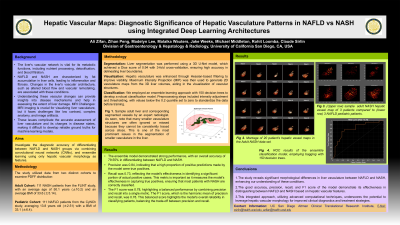
Introduction: Liver vasculature plays a pivotal role in metabolic function and disease progression, particularly in Non-alcoholic Fatty Liver Disease (NAFLD) and its more severe form, Non-Alcoholic Steatohepatitis (NASH). Challenges such as low MRI contrast, complex anatomy, and image artifacts hinder the development of accurate ground truths for training advanced machine learning models. Our study integrates traditional machine learning techniques with convolutional neural networks (CNNs) to differentiate between NAFLD and NASH, aiming to enhance diagnostic accuracy and refine treatment strategies based on hepatic vascular morphology.
Methods: We analyzed data from 227 patients, comprising 114 from the FLINT study (mean age: 51.2 ±11.5; BMI: 33.6±5.14) and 113 from the CyNCh study (age: 13.7±11.5; BMI: 32±6.8). Our methodology began with 3D liver segmentation using a 3D U-Net model, achieving a Dice score of 0.94 with 3-fold cross-validation. Subsequently, Hessian-based filtering enhanced hepatic vasculature visualization, followed by Maximum Intensity Projection (MIP) to generate 2D vasculature map for each patient from the 3D anatomical liver volumes. For classification, we employed ensemble learning methods, specifically bagging with 150 decision trees, to develop a robust model. Prior to training, preprocessing involved intensity adjustment and thresholding on the MIP images, setting all values below the 0.2 quantile to zero in both datasets, using a 3-fold cross-validation strategy.
Results: The ensemble model demonstrated promising predictive performance with an accuracy of 79.55%. Precision, measuring the proportion of true positive predictions among all positive predictions made by the model, was notably high at 0.84. The model also achieved a recall of 0.73, indicating its ability to correctly identify a substantial portion of actual positives. The F1 score, a harmonic mean of precision and recall, further validated the balanced performance of the ensemble model, yielding a score of 0.78.
Discussion: results underscore significant morphological differences in vasculature between NAFLD and NASH groups. The high accuracy, precision, recall, and F1 score collectively highlight the model's capability to discern these differences. This integrated approach emphasizes the utility of advanced computational methods in leveraging hepatic vascular morphology for enhanced clinical outcomes.

Disclosures:
Ali Zifan, PhD1, Zihan Peng, BSc1, Madilyn Lee, 1, Malaika Wauters, 1, Jake Weeks, BS1, Michael Middleton, PhD1, Rohit Loomba, MD, MHSc2, Claude Sirlin, MD1. P2854 - Hepatic Vascular Maps: Diagnostic Significance of Hepatic Vasculature Patterns in NAFLD vs NASH Using Integrated Deep Learning Architectures, ACG 2024 Annual Scientific Meeting Abstracts. Philadelphia, PA: American College of Gastroenterology.
1UC San Diego, La Jolla, CA; 2University of California San Diego School of Medicine, La Jolla, CA
Introduction: Liver vasculature plays a pivotal role in metabolic function and disease progression, particularly in Non-alcoholic Fatty Liver Disease (NAFLD) and its more severe form, Non-Alcoholic Steatohepatitis (NASH). Challenges such as low MRI contrast, complex anatomy, and image artifacts hinder the development of accurate ground truths for training advanced machine learning models. Our study integrates traditional machine learning techniques with convolutional neural networks (CNNs) to differentiate between NAFLD and NASH, aiming to enhance diagnostic accuracy and refine treatment strategies based on hepatic vascular morphology.
Methods: We analyzed data from 227 patients, comprising 114 from the FLINT study (mean age: 51.2 ±11.5; BMI: 33.6±5.14) and 113 from the CyNCh study (age: 13.7±11.5; BMI: 32±6.8). Our methodology began with 3D liver segmentation using a 3D U-Net model, achieving a Dice score of 0.94 with 3-fold cross-validation. Subsequently, Hessian-based filtering enhanced hepatic vasculature visualization, followed by Maximum Intensity Projection (MIP) to generate 2D vasculature map for each patient from the 3D anatomical liver volumes. For classification, we employed ensemble learning methods, specifically bagging with 150 decision trees, to develop a robust model. Prior to training, preprocessing involved intensity adjustment and thresholding on the MIP images, setting all values below the 0.2 quantile to zero in both datasets, using a 3-fold cross-validation strategy.
Results: The ensemble model demonstrated promising predictive performance with an accuracy of 79.55%. Precision, measuring the proportion of true positive predictions among all positive predictions made by the model, was notably high at 0.84. The model also achieved a recall of 0.73, indicating its ability to correctly identify a substantial portion of actual positives. The F1 score, a harmonic mean of precision and recall, further validated the balanced performance of the ensemble model, yielding a score of 0.78.
Discussion: results underscore significant morphological differences in vasculature between NAFLD and NASH groups. The high accuracy, precision, recall, and F1 score collectively highlight the model's capability to discern these differences. This integrated approach emphasizes the utility of advanced computational methods in leveraging hepatic vascular morphology for enhanced clinical outcomes.

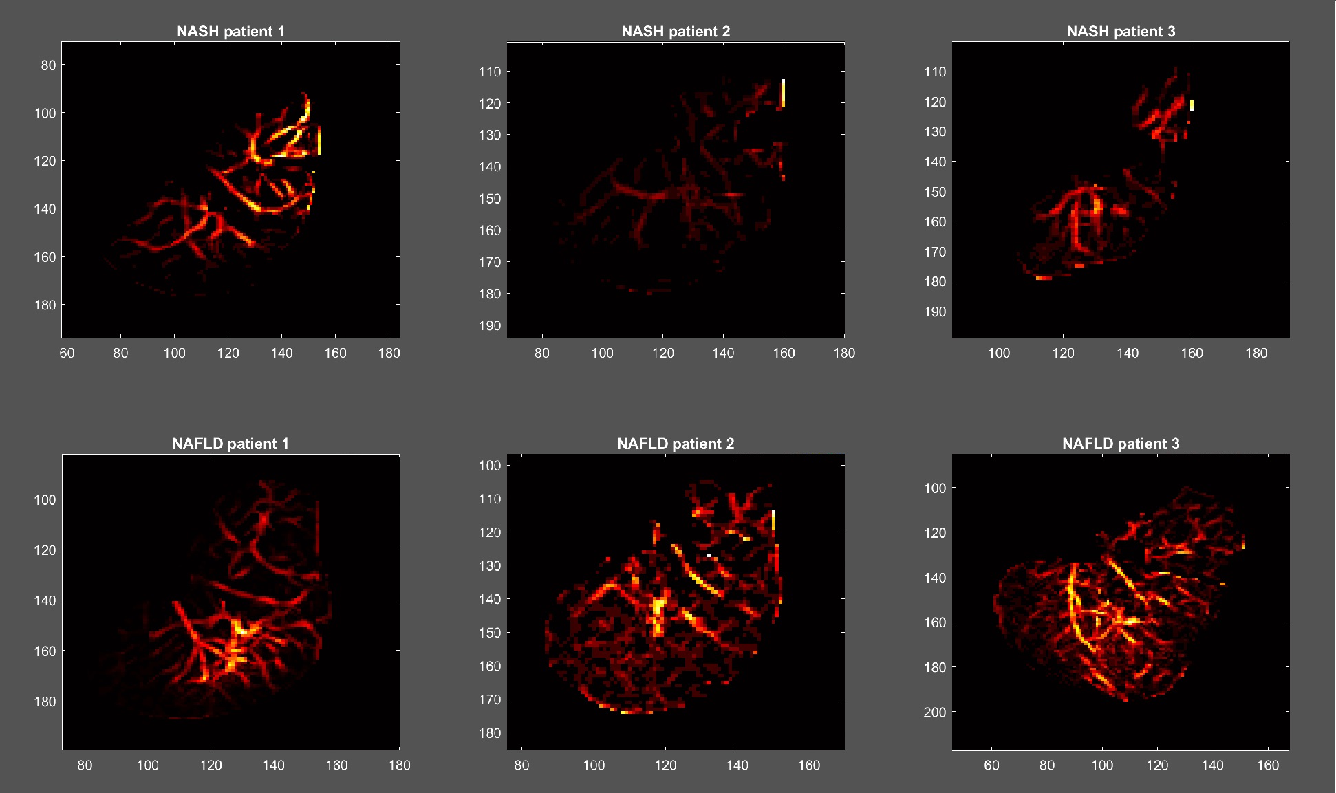
Figure: Figure 1. Hepatic vascular maps of pediatric NAFLD and adult Nash in 3 sample patients.
Disclosures:
Ali Zifan indicated no relevant financial relationships.
Zihan Peng indicated no relevant financial relationships.
Madilyn Lee indicated no relevant financial relationships.
Malaika Wauters indicated no relevant financial relationships.
Jake Weeks indicated no relevant financial relationships.
Michael Middleton indicated no relevant financial relationships.
Rohit Loomba: 89BIO – Consultant. Aardvark – Consultant. Altimmune – Consultant. Madrigal Pharmaceuticals – Consultant. Merck – Consultant. Novo Nordisk – Consultant. Takeda – Consultant. Terns – Consultant. Viking – Consultant.
Claude Sirlin indicated no relevant financial relationships.
Ali Zifan, PhD1, Zihan Peng, BSc1, Madilyn Lee, 1, Malaika Wauters, 1, Jake Weeks, BS1, Michael Middleton, PhD1, Rohit Loomba, MD, MHSc2, Claude Sirlin, MD1. P2854 - Hepatic Vascular Maps: Diagnostic Significance of Hepatic Vasculature Patterns in NAFLD vs NASH Using Integrated Deep Learning Architectures, ACG 2024 Annual Scientific Meeting Abstracts. Philadelphia, PA: American College of Gastroenterology.
