Monday Poster Session
Category: Interventional Endoscopy
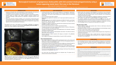
P2805 - Nonsurgical Treatment of Gangrenous Cholecystitis With EUS-Assisted Cholecystogastrostomy Using a Lumen-Apposing Metal Stent: First Case in the Literature
Monday, October 28, 2024
10:30 AM - 4:00 PM ET
Location: Exhibit Hall E

Has Audio
- MG
Mirac Gunay, MD
Morristown Medical Center
Morristown, NJ
Presenting Author(s)
Mirac Gunay, MD, Matthew Grossman, MD
Morristown Medical Center, Morristown, NJ
Introduction: Acute cholecystitis (AC) is the acute inflammation of the gallbladder and if left untreated, it can be complicated by gangrenous cholecystitis (GC). The definitive treatment of GC is cholecystectomy and although various alternative treatment modalities, including percutaneous cholecystostomy (PC) and EUS-guided drainage of gallbladder (EUS-GBD) using self-expanding metal stent (SEMS), are proposed and applied for patients with AC who are at high-risk for surgery, these alternative options for GC are scarcely described in the literature. To the best of our knowledge, there are no published cases for the drainage of gallbladder using lumen-apposing metal stents (LAMS) in GC.
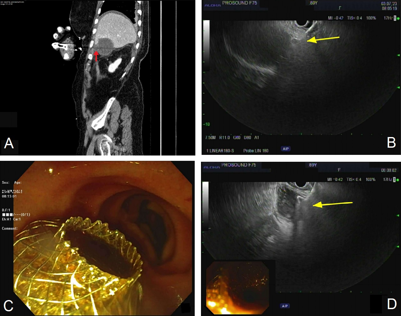
Case Description/Methods: An 89-year-old female presented to the hospital with 1-day history of diffuse abdominal pain, vomiting and worsening mental status. CT showed abnormally distended gallbladder (GB) with diffuse GB wall thickening, wall irregularity, and marked pericholecystic induration along with suspected perforation (Figure 1A). She was found to be a poor surgical candidate and underwent EUS-guided cholecystogastrotomy. Extensive hyperechoic material consistent with sludge was visualized in the gallbladder endosonographically. Subsequently, color Doppler imaging was utilized to identify superimposed vessels on the common wall between the stomach and the GB in order to find the most appropriate position in the stomach to create a cholecystogastrostomy. An electrocautery-enhanced LAMS was introduced through the working channel, current was applied to the cautery top and the ostomy was created. The LAMS device was advanced into the gallbladder and a 10x10 mm LAMS was placed with the flanges in close approximation to the walls of the gallbladder and the stomach through the cholecystogastrostomy. The stent was successfully placed with immediate drainage of dark brown fluid content and decompression of the gallbladder (Figure 1B, 1C, 1D). Her mental status and symptoms improved over subsequent days.
Discussion: Recent publications demonstrated lower rates of adverse events and recurrent cholecystitis in EUS-GBD using electrocautery-enhanced LAMS when compared to percutaneous drainage for treatment of AC. The literature suggests that PC can be used as a temporary measure for AC, however, it is advised to avoid PC in gangrenous cholecystitis likely due to fragile gallbladder wall secondary to necrosis. In such challenging situations, EUS-GBD using LAMS can likely be a safer option for the management of GC.

Disclosures:
Mirac Gunay, MD, Matthew Grossman, MD. P2805 - Nonsurgical Treatment of Gangrenous Cholecystitis With EUS-Assisted Cholecystogastrostomy Using a Lumen-Apposing Metal Stent: First Case in the Literature, ACG 2024 Annual Scientific Meeting Abstracts. Philadelphia, PA: American College of Gastroenterology.
Morristown Medical Center, Morristown, NJ
Introduction: Acute cholecystitis (AC) is the acute inflammation of the gallbladder and if left untreated, it can be complicated by gangrenous cholecystitis (GC). The definitive treatment of GC is cholecystectomy and although various alternative treatment modalities, including percutaneous cholecystostomy (PC) and EUS-guided drainage of gallbladder (EUS-GBD) using self-expanding metal stent (SEMS), are proposed and applied for patients with AC who are at high-risk for surgery, these alternative options for GC are scarcely described in the literature. To the best of our knowledge, there are no published cases for the drainage of gallbladder using lumen-apposing metal stents (LAMS) in GC.
Case Description/Methods: An 89-year-old female presented to the hospital with 1-day history of diffuse abdominal pain, vomiting and worsening mental status. CT showed abnormally distended gallbladder (GB) with diffuse GB wall thickening, wall irregularity, and marked pericholecystic induration along with suspected perforation (Figure 1A). She was found to be a poor surgical candidate and underwent EUS-guided cholecystogastrotomy. Extensive hyperechoic material consistent with sludge was visualized in the gallbladder endosonographically. Subsequently, color Doppler imaging was utilized to identify superimposed vessels on the common wall between the stomach and the GB in order to find the most appropriate position in the stomach to create a cholecystogastrostomy. An electrocautery-enhanced LAMS was introduced through the working channel, current was applied to the cautery top and the ostomy was created. The LAMS device was advanced into the gallbladder and a 10x10 mm LAMS was placed with the flanges in close approximation to the walls of the gallbladder and the stomach through the cholecystogastrostomy. The stent was successfully placed with immediate drainage of dark brown fluid content and decompression of the gallbladder (Figure 1B, 1C, 1D). Her mental status and symptoms improved over subsequent days.
Discussion: Recent publications demonstrated lower rates of adverse events and recurrent cholecystitis in EUS-GBD using electrocautery-enhanced LAMS when compared to percutaneous drainage for treatment of AC. The literature suggests that PC can be used as a temporary measure for AC, however, it is advised to avoid PC in gangrenous cholecystitis likely due to fragile gallbladder wall secondary to necrosis. In such challenging situations, EUS-GBD using LAMS can likely be a safer option for the management of GC.

Figure: Figure 1. (A) Areas of irregularity of the gallbladder wall with suspected perforation (red arrow). (B) EUS showing deployment of LAMS between the gallbladder and stomach wall (yellow arrow). (C) Endoscopic view of the LAMS placed through the cholecystogastrostomy. (D) EUS showing the decompressed gallbladder following the placement of the LAMS (yellow arrow).
Disclosures:
Mirac Gunay indicated no relevant financial relationships.
Matthew Grossman indicated no relevant financial relationships.
Mirac Gunay, MD, Matthew Grossman, MD. P2805 - Nonsurgical Treatment of Gangrenous Cholecystitis With EUS-Assisted Cholecystogastrostomy Using a Lumen-Apposing Metal Stent: First Case in the Literature, ACG 2024 Annual Scientific Meeting Abstracts. Philadelphia, PA: American College of Gastroenterology.
