Monday Poster Session
Category: General Endoscopy
P2439 - Diagnosis of Trichuriasis by Colonoscopy
Monday, October 28, 2024
10:30 AM - 4:00 PM ET
Location: Exhibit Hall E

Has Audio
- NS
Nancy Seto, MS, DO
Kirk Kerkorian School of Medicine at the University of Nevada
Las Vegas, NV
Presenting Author(s)
Nancy Seto, MS, DO, Preet Patel, MD, Gordon Ohning, MD, PhD
Kirk Kerkorian School of Medicine at the University of Nevada, Las Vegas, NV
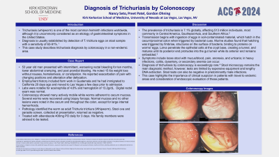
Introduction: Trichuriasis (whipworm) is one of the most common helminth infections worldwide, although in the US, it is uncommonly considered as an etiology of gastrointestinal symptoms. Diagnosis is usually established by detection of T. trichiura eggs on stool sample examination with a sensitivity of 63-91%. This case study describes trichuriasis diagnosis by colonoscopy in a non-endemic area.
Case Description/Methods: A 53 year old man presented with intermittent, worsening rectal bleeding for ten months, lower abdominal cramping, and post prandial bloating. He noted 15 lbs weight loss without nausea, hematemesis, or constipation. He reported exacerbation of pain with changing positions and alleviation after defecation. His employment history included farm work in Guatemala and he had immigrated to California 20 days ago and moved to Las Vegas a few days prior to admission.
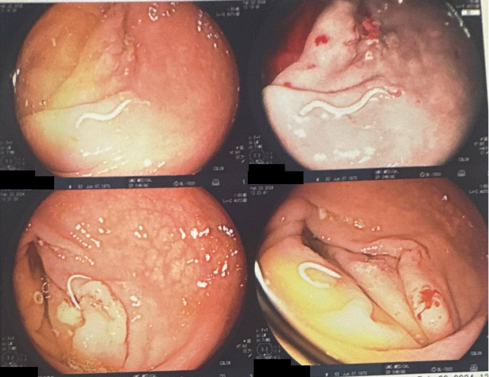
Labs were notable for eosinophilia of 4.8% and hemoglobin of 13.2g/dL. Digital rectal exam was normal. He underwent colonoscopy and was found to have many actively mobile white worms adherent to cecum mucosa. Several worms were recovered using biopsy forceps. Normal mucosa and no other lesions were noted in the cecum and throughout the colon, except for large internal hemorrhoids. Pathology identified the worm as adult Trichuris trichiura (Whipworm). Stool ova and parasite returned as negative.
The patient was treated with albendazole 400mg PO daily for 3 days. His family members were advised to be tested.
Discussion: The prevalence of trichuriasis is 7.1% globally, affecting 513 million individuals, most commonly in Central America, Southeast Asia, and Southern Africa. Transmission begins with ingestion of eggs in soil-contaminated material, which hatch in the cecum/proximal colon when triggered by bacterial cues. Larva penetrate the epithelial cells at the crypt base, creating a tunnel, and matures until its posterior end protrudes into the gut lumen. Symptoms include loose stool with mucus/blood, pain, anorexia, and urticaria; in heavy infections, colitis, dysentery, or secondary anemia can occur. To our knowledge, diagnosis of trichuriasis by colonoscopy is exceedingly rare. Stool microscopy remains the main diagnostic method, however, tests are limited by expensive equipment and lengthy DNA extraction. Stool tests can also be negative in predominantly male infections. This case highlights the importance of clinical suspicion in patients with travel to endemic areas and consideration of endoscopic evaluation of these patients.

Disclosures:
Nancy Seto, MS, DO, Preet Patel, MD, Gordon Ohning, MD, PhD. P2439 - Diagnosis of Trichuriasis by Colonoscopy, ACG 2024 Annual Scientific Meeting Abstracts. Philadelphia, PA: American College of Gastroenterology.
Kirk Kerkorian School of Medicine at the University of Nevada, Las Vegas, NV
Introduction: Trichuriasis (whipworm) is one of the most common helminth infections worldwide, although in the US, it is uncommonly considered as an etiology of gastrointestinal symptoms. Diagnosis is usually established by detection of T. trichiura eggs on stool sample examination with a sensitivity of 63-91%. This case study describes trichuriasis diagnosis by colonoscopy in a non-endemic area.
Case Description/Methods: A 53 year old man presented with intermittent, worsening rectal bleeding for ten months, lower abdominal cramping, and post prandial bloating. He noted 15 lbs weight loss without nausea, hematemesis, or constipation. He reported exacerbation of pain with changing positions and alleviation after defecation. His employment history included farm work in Guatemala and he had immigrated to California 20 days ago and moved to Las Vegas a few days prior to admission.
Labs were notable for eosinophilia of 4.8% and hemoglobin of 13.2g/dL. Digital rectal exam was normal. He underwent colonoscopy and was found to have many actively mobile white worms adherent to cecum mucosa. Several worms were recovered using biopsy forceps. Normal mucosa and no other lesions were noted in the cecum and throughout the colon, except for large internal hemorrhoids. Pathology identified the worm as adult Trichuris trichiura (Whipworm). Stool ova and parasite returned as negative.
The patient was treated with albendazole 400mg PO daily for 3 days. His family members were advised to be tested.
Discussion: The prevalence of trichuriasis is 7.1% globally, affecting 513 million individuals, most commonly in Central America, Southeast Asia, and Southern Africa. Transmission begins with ingestion of eggs in soil-contaminated material, which hatch in the cecum/proximal colon when triggered by bacterial cues. Larva penetrate the epithelial cells at the crypt base, creating a tunnel, and matures until its posterior end protrudes into the gut lumen. Symptoms include loose stool with mucus/blood, pain, anorexia, and urticaria; in heavy infections, colitis, dysentery, or secondary anemia can occur. To our knowledge, diagnosis of trichuriasis by colonoscopy is exceedingly rare. Stool microscopy remains the main diagnostic method, however, tests are limited by expensive equipment and lengthy DNA extraction. Stool tests can also be negative in predominantly male infections. This case highlights the importance of clinical suspicion in patients with travel to endemic areas and consideration of endoscopic evaluation of these patients.

Figure: Adult Trichuris trichiura adherent to cecum mucosa on colonoscopy
Disclosures:
Nancy Seto indicated no relevant financial relationships.
Preet Patel indicated no relevant financial relationships.
Gordon Ohning indicated no relevant financial relationships.
Nancy Seto, MS, DO, Preet Patel, MD, Gordon Ohning, MD, PhD. P2439 - Diagnosis of Trichuriasis by Colonoscopy, ACG 2024 Annual Scientific Meeting Abstracts. Philadelphia, PA: American College of Gastroenterology.
