Monday Poster Session
Category: Esophagus
P2305 - The Emerging Role of Endoscopic Ultrasound in Diagnosing Dysphagia Lusoria
Monday, October 28, 2024
10:30 AM - 4:00 PM ET
Location: Exhibit Hall E

Has Audio

Aleem A. Ali, MBBS
University of Florida Health Shands Hospital
Jacksonville, FL
Presenting Author(s)
Aleem A. Ali, MBBS1, Oshin Rai, DO2, Aarti Maharaj, MD1, Bruno Ribeiro, MD2
1University of Florida Health Shands Hospital, Jacksonville, FL; 2University of Florida College of Medicine, Jacksonville, FL
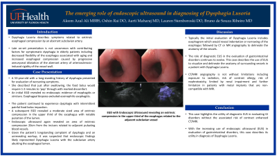
Introduction: Dysphagia lusoria, resulting from the external compression of the esophagus by an aberrant right subclavian artery (ARSA), is rare, with a prevalence of less than 1%. Symptomatic patients with ARSA are typically around 48 years old, influenced by factors such as age-related decline in esophageal flexibility and increased compression from progressive aneurysmal dilation or arteriosclerosis-induced vessel wall rigidity. Initial assessment using an esophagram often reveals esophageal indentation or narrowing, further elucidated by CT or MR angiography to assess the vascular anatomy. This case underscores the expanding role of endoscopic ultrasound (EUS) in diagnosing Dysphagia Lusoria.
Case Description/Methods: A 53-year-old woman with a longstanding history of dysphagia presented for evaluation of progressively worsening symptoms. She described significant discomfort, noting that it took 1-2 minutes for the food bolus to pass into her stomach after swallowing. She underwent esophagogastroduodenoscopy (EGD), which ruled out esophagitis or strictures, and esophageal biopsies confirmed the absence of eosinophilic esophagitis. She continued to experience dysphagia with intermittent painful food bolus impaction. She described these episodes as a sensation of food being 'encapsulated in a bubble' in her neck.
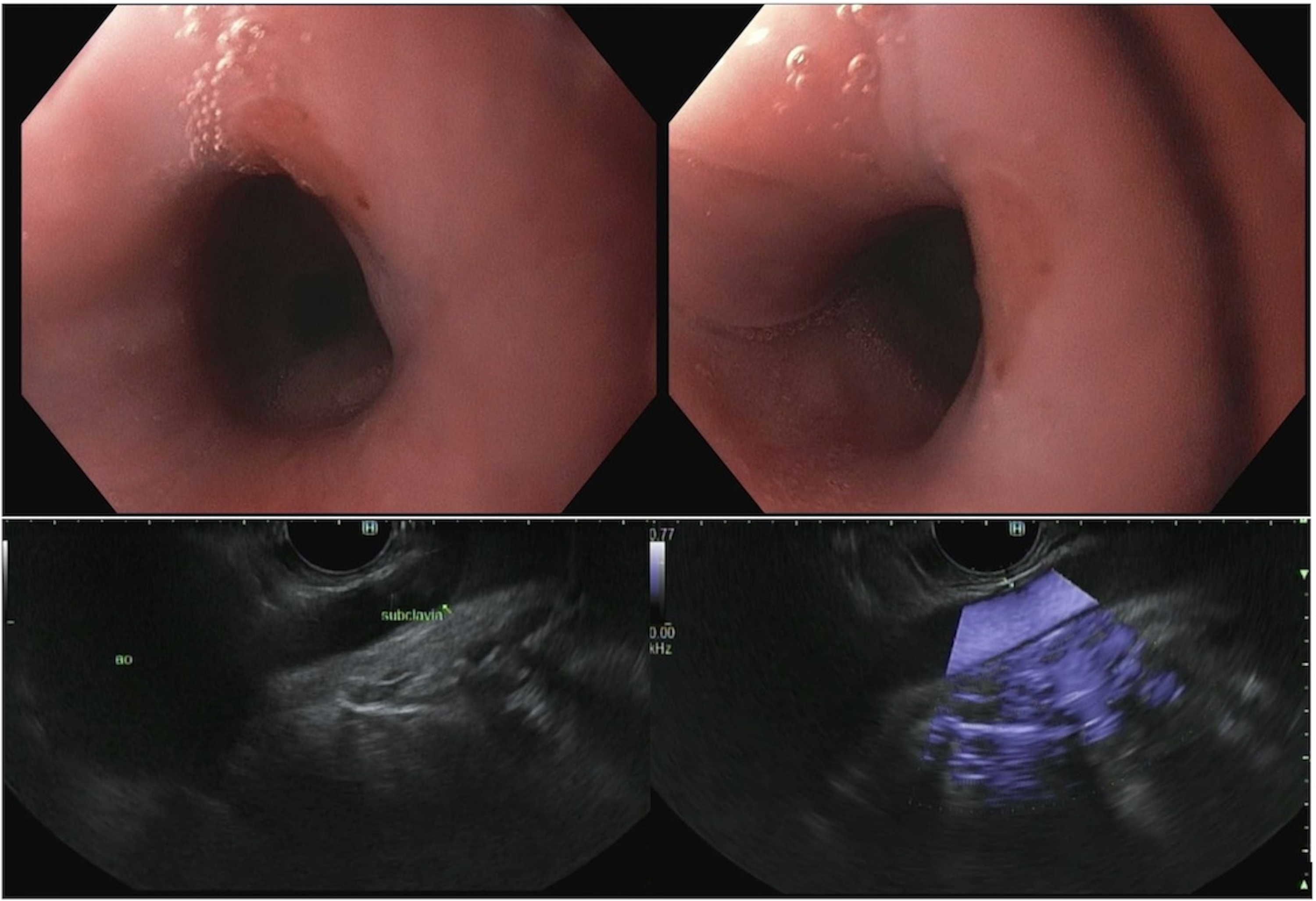
A subsequent EGD identified a moderate-sized area of extrinsic compression in the upper third of the esophagus, notable for pulsation within the lumen. Endoscopic ultrasound confirmed an area of extrinsic compression 20cm from the incisors, associated with nearby pulsating blood vessels. Considering the patient’s longstanding dysphagia and unrevealing prior evaluations, these findings raised high suspicion for dysphagia lusoria.
Discussion: The role of diagnostic EUS in the evaluation of gastrointestinal disorders continues to evolve. This case illustrates its use in visualizing and mapping surrounding vessel anatomy in a patient with dysphagia lusoria. CT/MRI angiography has limitations, including radiation exposure, risk of contrast allergy, nephrotoxicity, and additional constraints for patients with non-MRI-compatible implanted devices. This case underscores the utility of EUS in assessing GI disorders without the risks associated with contrast-enhanced imaging modalities.

Disclosures:
Aleem A. Ali, MBBS1, Oshin Rai, DO2, Aarti Maharaj, MD1, Bruno Ribeiro, MD2. P2305 - The Emerging Role of Endoscopic Ultrasound in Diagnosing Dysphagia Lusoria, ACG 2024 Annual Scientific Meeting Abstracts. Philadelphia, PA: American College of Gastroenterology.
1University of Florida Health Shands Hospital, Jacksonville, FL; 2University of Florida College of Medicine, Jacksonville, FL
Introduction: Dysphagia lusoria, resulting from the external compression of the esophagus by an aberrant right subclavian artery (ARSA), is rare, with a prevalence of less than 1%. Symptomatic patients with ARSA are typically around 48 years old, influenced by factors such as age-related decline in esophageal flexibility and increased compression from progressive aneurysmal dilation or arteriosclerosis-induced vessel wall rigidity. Initial assessment using an esophagram often reveals esophageal indentation or narrowing, further elucidated by CT or MR angiography to assess the vascular anatomy. This case underscores the expanding role of endoscopic ultrasound (EUS) in diagnosing Dysphagia Lusoria.
Case Description/Methods: A 53-year-old woman with a longstanding history of dysphagia presented for evaluation of progressively worsening symptoms. She described significant discomfort, noting that it took 1-2 minutes for the food bolus to pass into her stomach after swallowing. She underwent esophagogastroduodenoscopy (EGD), which ruled out esophagitis or strictures, and esophageal biopsies confirmed the absence of eosinophilic esophagitis. She continued to experience dysphagia with intermittent painful food bolus impaction. She described these episodes as a sensation of food being 'encapsulated in a bubble' in her neck.
A subsequent EGD identified a moderate-sized area of extrinsic compression in the upper third of the esophagus, notable for pulsation within the lumen. Endoscopic ultrasound confirmed an area of extrinsic compression 20cm from the incisors, associated with nearby pulsating blood vessels. Considering the patient’s longstanding dysphagia and unrevealing prior evaluations, these findings raised high suspicion for dysphagia lusoria.
Discussion: The role of diagnostic EUS in the evaluation of gastrointestinal disorders continues to evolve. This case illustrates its use in visualizing and mapping surrounding vessel anatomy in a patient with dysphagia lusoria. CT/MRI angiography has limitations, including radiation exposure, risk of contrast allergy, nephrotoxicity, and additional constraints for patients with non-MRI-compatible implanted devices. This case underscores the utility of EUS in assessing GI disorders without the risks associated with contrast-enhanced imaging modalities.

Figure: Endosonographic findings of esophageal narrowing with the subclavian artery just abutting the oesophageal lumen.
Disclosures:
Aleem Ali indicated no relevant financial relationships.
Oshin Rai indicated no relevant financial relationships.
Aarti Maharaj indicated no relevant financial relationships.
Bruno Ribeiro indicated no relevant financial relationships.
Aleem A. Ali, MBBS1, Oshin Rai, DO2, Aarti Maharaj, MD1, Bruno Ribeiro, MD2. P2305 - The Emerging Role of Endoscopic Ultrasound in Diagnosing Dysphagia Lusoria, ACG 2024 Annual Scientific Meeting Abstracts. Philadelphia, PA: American College of Gastroenterology.

