Monday Poster Session
Category: Esophagus
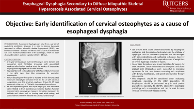
P2307 - Esophageal Dysphagia Secondary to Diffuse Idiopathic Skeletal Hyperostosis-Associated Cervical Osteophytes
Monday, October 28, 2024
10:30 AM - 4:00 PM ET
Location: Exhibit Hall E

Has Audio
.jpg)
Arvind Bussetty, MD
Robert Wood Johnson Medical School, Rutgers University, NJ
Presenting Author(s)
Arvind Bussetty, MD, Anish V. Patel, MD
Robert Wood Johnson Medical School, Rutgers University, New Brunswick, NJ
Introduction: Esophageal Dysphagia can arise from a variety of underlying conditions. However, it is rare to observe dysphagia secondary to diffuse idiopathic skeletal hyperostosis (DISH), also known as Forestier’s disease. The excrescence of cervical osteophytes can cause mechanical obstruction of the esophagus, which has been observed in elderly patients typically in the C4-C5 level.
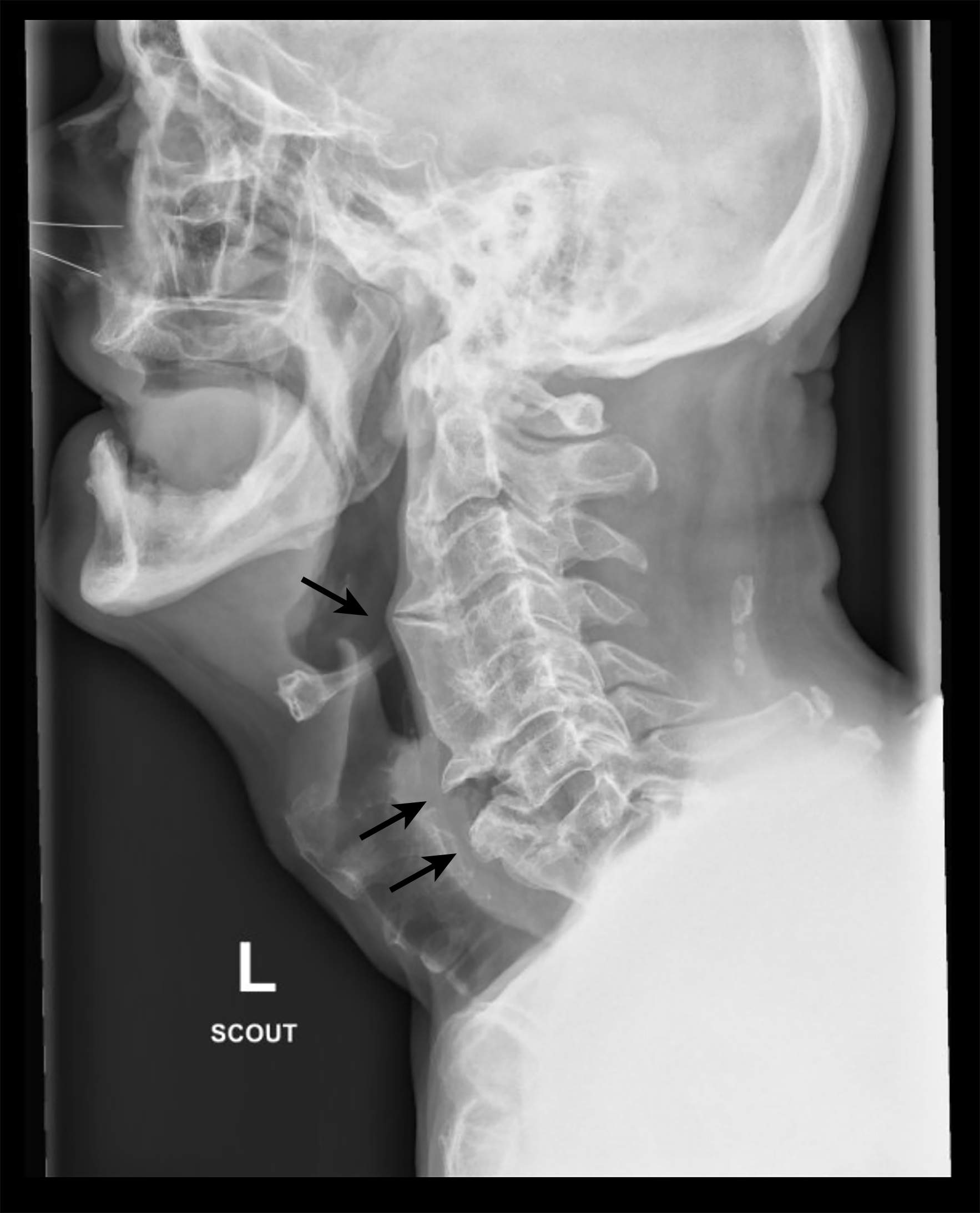
Case Description/Methods: A 78-year-old Caucasian male with a past medical history of aortic stenosis and paroxysmal atrial fibrillation presented with generalized weakness after barium swallow study with an outside gastroenterology for workup of progressive dysphagia and regurgitation for three months. Patient presented with leukocytosis and chest X-ray showed barium material in the right lower lung lobe concerning for aspiration pneumonitis. Barium Esophagram performed before hospital arrival demonstrated exuberant osteophyte formation from C3 to C7 with extrinsic compression onto the proximal esophagus (Figure 1). CT of the neck supported a diagnosis of DISH with large cervical osteophytes. Patient was experiencing solid and liquid dysphagia, and antibiotics were initiated to treat aspiration pneumonia. Swallow function improved with conservative measures including maneuvers to facilitate oral intake such as turning head while eating, and gradual advancement of diet.
Discussion: We present here a case of DISH discovered by esophagram evaluation found to be the etiology of dysphagia. Mild to moderate symptoms can be managed with pain medications and swallowing techniques. Osteophyte resection may be required in cases of weight loss or severe dysphagia to solids or liquids. In our case, the patient was a poor candidate for surgical treatment. However, conservative measures with pain control and gradual diet introduction were successful in managing symptoms for this case. Patient counseling and compliance with dietary modification, and speech and swallow therapy are essential. This diagnosis should be considered when evaluating dysphagia, especially in the older population. XR Esophagram should be performed early in such patient populations especially to identify obstructive esophageal pathology such as osteophytes, and can be used for non-invasive surveillance of disease severity.

Disclosures:
Arvind Bussetty, MD, Anish V. Patel, MD. P2307 - Esophageal Dysphagia Secondary to Diffuse Idiopathic Skeletal Hyperostosis-Associated Cervical Osteophytes, ACG 2024 Annual Scientific Meeting Abstracts. Philadelphia, PA: American College of Gastroenterology.
Robert Wood Johnson Medical School, Rutgers University, New Brunswick, NJ
Introduction: Esophageal Dysphagia can arise from a variety of underlying conditions. However, it is rare to observe dysphagia secondary to diffuse idiopathic skeletal hyperostosis (DISH), also known as Forestier’s disease. The excrescence of cervical osteophytes can cause mechanical obstruction of the esophagus, which has been observed in elderly patients typically in the C4-C5 level.
Case Description/Methods: A 78-year-old Caucasian male with a past medical history of aortic stenosis and paroxysmal atrial fibrillation presented with generalized weakness after barium swallow study with an outside gastroenterology for workup of progressive dysphagia and regurgitation for three months. Patient presented with leukocytosis and chest X-ray showed barium material in the right lower lung lobe concerning for aspiration pneumonitis. Barium Esophagram performed before hospital arrival demonstrated exuberant osteophyte formation from C3 to C7 with extrinsic compression onto the proximal esophagus (Figure 1). CT of the neck supported a diagnosis of DISH with large cervical osteophytes. Patient was experiencing solid and liquid dysphagia, and antibiotics were initiated to treat aspiration pneumonia. Swallow function improved with conservative measures including maneuvers to facilitate oral intake such as turning head while eating, and gradual advancement of diet.
Discussion: We present here a case of DISH discovered by esophagram evaluation found to be the etiology of dysphagia. Mild to moderate symptoms can be managed with pain medications and swallowing techniques. Osteophyte resection may be required in cases of weight loss or severe dysphagia to solids or liquids. In our case, the patient was a poor candidate for surgical treatment. However, conservative measures with pain control and gradual diet introduction were successful in managing symptoms for this case. Patient counseling and compliance with dietary modification, and speech and swallow therapy are essential. This diagnosis should be considered when evaluating dysphagia, especially in the older population. XR Esophagram should be performed early in such patient populations especially to identify obstructive esophageal pathology such as osteophytes, and can be used for non-invasive surveillance of disease severity.

Figure: 1: Barium esophagram demonstrating an exuberant osteophyte leading to esophageal compression and dysphagia.
Disclosures:
Arvind Bussetty indicated no relevant financial relationships.
Anish Patel indicated no relevant financial relationships.
Arvind Bussetty, MD, Anish V. Patel, MD. P2307 - Esophageal Dysphagia Secondary to Diffuse Idiopathic Skeletal Hyperostosis-Associated Cervical Osteophytes, ACG 2024 Annual Scientific Meeting Abstracts. Philadelphia, PA: American College of Gastroenterology.
