Monday Poster Session
Category: Colon
P2065 - An Unexpected Detour: Partial Large Bowel Obstruction Due to Rectal Carcinoma in a Young Adult
Monday, October 28, 2024
10:30 AM - 4:00 PM ET
Location: Exhibit Hall E

Has Audio
- SS
Sonia Samuel, DO
Albany Medical Center
Albany, NY
Presenting Author(s)
Sonia Samuel, DO, Ahmad Abulawi, MD, Asra Batool, MD
Albany Medical Center, Albany, NY
Introduction: The incidence of colorectal cancer in young adults is rising worldwide. In those under the age of 50, it is the first and second leading causes of death in men and women, respectively. Symptoms vary but can initially present with a large bowel obstruction. We present a case of a young female who develops a partial large bowel obstruction due to rectal carcinoma.
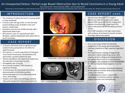
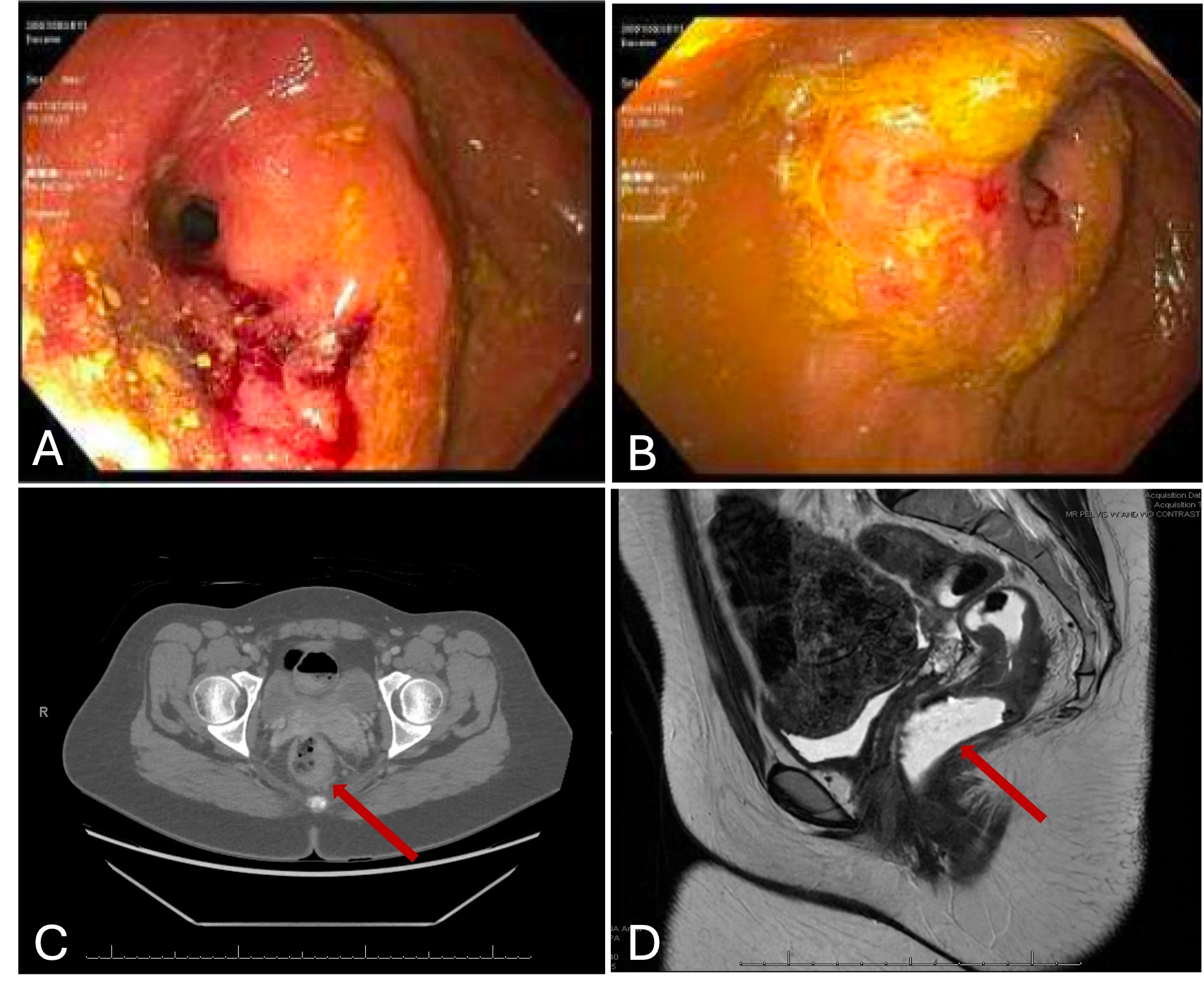
Case Description/Methods: A 24-year-old female with no significant past medical history presented to the office for abdominal pain. She reports a 1-month long history of frequent loose bowel movements, hematochezia and generalized, constant abdominal pain. On physical exam, patient had diffuse mild abdominal tenderness, but soft with normal bowel sounds. Hemoglobin was 13.0 g/dL. At an outside hospital, computed tomography (CT) of the abdomen and pelvis showed no significant findings. A colonoscopy was then performed which demonstrated an infiltrative, partially obstructing large 6 cm circumferential rectal mass, with oozing (Figure 1A and 1B). Biopsies were taken and pathology revealed high grade dysplasia/intramucosal carcinoma. Carcinoembryonic antigen (CEA) level was elevated at 17.3 ng/mL. Patient was referred to colorectal surgery. Repeat sigmoidoscopy by colorectal surgery demonstrated the similar fungating, ulcerative mass at the mid-rectum. Repeat CT Abdomen and Pelvis was performed for staging and showed circumferential thickening of the rectum with peri-rectal fat stranding and stool burden (Figure 1C). CT Chest showed no metastatic lesions. Pelvic MRI revealed a mid-high rectal tumor with evidence of extramural vascular invasion (Figure 1D). Oncology referral was placed for further treatment options.
Discussion: Due to the rising number of colorectal malignancy in the young adult population, clinicians should be of high suspicion regardless of age. The most common etiology of large bowel obstruction (LBO) is colorectal cancer, with majority of obstructing lesions being distal to the splenic flexure due to its narrow lumen. CT imaging is the modality of choice to evaluate for LBO but may be falsely negative in partial LBO. The need for immediate intervention depends on the severity of bowel obstruction including concern for ischemia. A multidisciplinary team is required for exploration of treatment options such as surgical resection and chemotherapy.

Disclosures:
Sonia Samuel, DO, Ahmad Abulawi, MD, Asra Batool, MD. P2065 - An Unexpected Detour: Partial Large Bowel Obstruction Due to Rectal Carcinoma in a Young Adult, ACG 2024 Annual Scientific Meeting Abstracts. Philadelphia, PA: American College of Gastroenterology.
Albany Medical Center, Albany, NY
Introduction: The incidence of colorectal cancer in young adults is rising worldwide. In those under the age of 50, it is the first and second leading causes of death in men and women, respectively. Symptoms vary but can initially present with a large bowel obstruction. We present a case of a young female who develops a partial large bowel obstruction due to rectal carcinoma.
Case Description/Methods: A 24-year-old female with no significant past medical history presented to the office for abdominal pain. She reports a 1-month long history of frequent loose bowel movements, hematochezia and generalized, constant abdominal pain. On physical exam, patient had diffuse mild abdominal tenderness, but soft with normal bowel sounds. Hemoglobin was 13.0 g/dL. At an outside hospital, computed tomography (CT) of the abdomen and pelvis showed no significant findings. A colonoscopy was then performed which demonstrated an infiltrative, partially obstructing large 6 cm circumferential rectal mass, with oozing (Figure 1A and 1B). Biopsies were taken and pathology revealed high grade dysplasia/intramucosal carcinoma. Carcinoembryonic antigen (CEA) level was elevated at 17.3 ng/mL. Patient was referred to colorectal surgery. Repeat sigmoidoscopy by colorectal surgery demonstrated the similar fungating, ulcerative mass at the mid-rectum. Repeat CT Abdomen and Pelvis was performed for staging and showed circumferential thickening of the rectum with peri-rectal fat stranding and stool burden (Figure 1C). CT Chest showed no metastatic lesions. Pelvic MRI revealed a mid-high rectal tumor with evidence of extramural vascular invasion (Figure 1D). Oncology referral was placed for further treatment options.
Discussion: Due to the rising number of colorectal malignancy in the young adult population, clinicians should be of high suspicion regardless of age. The most common etiology of large bowel obstruction (LBO) is colorectal cancer, with majority of obstructing lesions being distal to the splenic flexure due to its narrow lumen. CT imaging is the modality of choice to evaluate for LBO but may be falsely negative in partial LBO. The need for immediate intervention depends on the severity of bowel obstruction including concern for ischemia. A multidisciplinary team is required for exploration of treatment options such as surgical resection and chemotherapy.

Figure: Figure 1A and 1B- Colonoscopy of rectal mass with oozing. 1C- CT Abdomen and Pelvis shows rectal thickening. 1D- Pelvic MRI shows a 6.2 cm rectal tumor
Disclosures:
Sonia Samuel indicated no relevant financial relationships.
Ahmad Abulawi indicated no relevant financial relationships.
Asra Batool indicated no relevant financial relationships.
Sonia Samuel, DO, Ahmad Abulawi, MD, Asra Batool, MD. P2065 - An Unexpected Detour: Partial Large Bowel Obstruction Due to Rectal Carcinoma in a Young Adult, ACG 2024 Annual Scientific Meeting Abstracts. Philadelphia, PA: American College of Gastroenterology.
