Monday Poster Session
Category: Colon
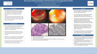
P2069 - Rare Case of Mucosal Schwann Cell Hamartoma in the Appendiceal Orifice
Monday, October 28, 2024
10:30 AM - 4:00 PM ET
Location: Exhibit Hall E

Has Audio
- BC
Brian H. Chung, MPH
Emory University
Lawrenceville, GA
Presenting Author(s)
Brian H. Chung, MPH1, Geetha Jagannathan, MD2, Jason Brown, MD3
1Emory University, Lawrenceville, GA; 2Emory University School of Medicine, Decatur, GA; 3Emory University School of Medicine, Atlanta, GA
Introduction: First identified in 2009, Mucosal Schwann Cell Hamartomas (MSCH) are rare, benign tumors originating from perineural Schwann cells found in the GI system. They are most found in the sigmoid colon and rarely found in the cecum, with only 10% of cases found there in a recent review.
We report a patient with a diminutive, sessile polyp in the appendiceal orifice found during a routine screening colonoscopy.
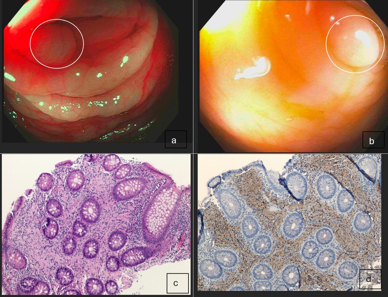
Case Description/Methods: The patient is a 58-year-old male with a history of alcohol misuse, alcohol-related pancreatitis, heartburn, and heart failure who presented to GI clinic after a pancreatic cyst was identified on imaging during an ED admission for acute pancreatitis. EUS-FNA was ordered for the pancreatic cyst, and colonoscopy was ordered upon learning of maternal history of colon cancer and no past colon cancer screening. A 2 mm sessile polyp was found in the appendiceal orifice and removed by cold forceps (1a & 1b). Pathology results showed bland spindle cell proliferation in the lamina propria of the colonic mucosa and S-100 staining returned positive, highlighting lesional spindle cells (1c & 1d).
The patient was counseled on the diagnosis of MSCH and advised to return in 3 years for surveillance colonoscopy due to his bowel prep quality.
Discussion: With colonoscopy volumes expected to rise due to guideline changes and demographic shifts, more MSCH are likely to be discovered. Therefore, it is important for providers to be aware of MSCH and understand how to distinguish it from other lesions.
Proper histology techniques can differentiate MSCH from other neural lesions such as schwannomas, neurofibromas, and ganglioneuromas. MSCH can be distinguished by proliferation of uniform spindle cells with unclear cell borders, dense eosinophilic cytoplasm, and elongated nuclei in the lamina propria. On immunohistochemistry, MSCH stain positive for S-100 protein and negative for other stains such as CD34, smooth muscle actin, epithelial membrane antigen, and NFP.
The location of this MSCH lesion in the appendiceal orifice was unique as lesions in this location have an overall prevalence of 0.08%.
This case illustrates a rare presentation of MSCH; moreover, the presence of an MSCH in the appendiceal orifice was unusual, as these lesions are thought to predominantly affect the distal colon. This case highlights the importance of including MSCH on the differential regardless of the area of presentation and working with pathologists who can accurately stain for and assess these lesions.

Disclosures:
Brian H. Chung, MPH1, Geetha Jagannathan, MD2, Jason Brown, MD3. P2069 - Rare Case of Mucosal Schwann Cell Hamartoma in the Appendiceal Orifice, ACG 2024 Annual Scientific Meeting Abstracts. Philadelphia, PA: American College of Gastroenterology.
1Emory University, Lawrenceville, GA; 2Emory University School of Medicine, Decatur, GA; 3Emory University School of Medicine, Atlanta, GA
Introduction: First identified in 2009, Mucosal Schwann Cell Hamartomas (MSCH) are rare, benign tumors originating from perineural Schwann cells found in the GI system. They are most found in the sigmoid colon and rarely found in the cecum, with only 10% of cases found there in a recent review.
We report a patient with a diminutive, sessile polyp in the appendiceal orifice found during a routine screening colonoscopy.
Case Description/Methods: The patient is a 58-year-old male with a history of alcohol misuse, alcohol-related pancreatitis, heartburn, and heart failure who presented to GI clinic after a pancreatic cyst was identified on imaging during an ED admission for acute pancreatitis. EUS-FNA was ordered for the pancreatic cyst, and colonoscopy was ordered upon learning of maternal history of colon cancer and no past colon cancer screening. A 2 mm sessile polyp was found in the appendiceal orifice and removed by cold forceps (1a & 1b). Pathology results showed bland spindle cell proliferation in the lamina propria of the colonic mucosa and S-100 staining returned positive, highlighting lesional spindle cells (1c & 1d).
The patient was counseled on the diagnosis of MSCH and advised to return in 3 years for surveillance colonoscopy due to his bowel prep quality.
Discussion: With colonoscopy volumes expected to rise due to guideline changes and demographic shifts, more MSCH are likely to be discovered. Therefore, it is important for providers to be aware of MSCH and understand how to distinguish it from other lesions.
Proper histology techniques can differentiate MSCH from other neural lesions such as schwannomas, neurofibromas, and ganglioneuromas. MSCH can be distinguished by proliferation of uniform spindle cells with unclear cell borders, dense eosinophilic cytoplasm, and elongated nuclei in the lamina propria. On immunohistochemistry, MSCH stain positive for S-100 protein and negative for other stains such as CD34, smooth muscle actin, epithelial membrane antigen, and NFP.
The location of this MSCH lesion in the appendiceal orifice was unique as lesions in this location have an overall prevalence of 0.08%.
This case illustrates a rare presentation of MSCH; moreover, the presence of an MSCH in the appendiceal orifice was unusual, as these lesions are thought to predominantly affect the distal colon. This case highlights the importance of including MSCH on the differential regardless of the area of presentation and working with pathologists who can accurately stain for and assess these lesions.

Figure: a. Narrowband imaging endo shot
b. Regular white light endo shot
c. H&E stain of colonic mucosa showing bland spindle cell proliferation in the lamina propria
d. Positive S-100 stain of lesional spindle cells
b. Regular white light endo shot
c. H&E stain of colonic mucosa showing bland spindle cell proliferation in the lamina propria
d. Positive S-100 stain of lesional spindle cells
Disclosures:
Brian Chung indicated no relevant financial relationships.
Geetha Jagannathan indicated no relevant financial relationships.
Jason Brown indicated no relevant financial relationships.
Brian H. Chung, MPH1, Geetha Jagannathan, MD2, Jason Brown, MD3. P2069 - Rare Case of Mucosal Schwann Cell Hamartoma in the Appendiceal Orifice, ACG 2024 Annual Scientific Meeting Abstracts. Philadelphia, PA: American College of Gastroenterology.

