Monday Poster Session
Category: Colon
P2007 - Lobular Capillary Hemangioma: A Rare Bleeding Entity in the GI Tract
Monday, October 28, 2024
10:30 AM - 4:00 PM ET
Location: Exhibit Hall E

Has Audio

Hammad Qadri, DO
United Health Services, Wilson Medical Center
Vestal, NY
Presenting Author(s)
Hammad Qadri, DO1, Yasir Ahmed, MD1, Nour Gafsi, BSc2, Usama Sakhawat, MD1, Amanke Oranu, MD1
1United Health Services, Wilson Medical Center, Binghamton, NY; 2United Health Services, Wilson Medical Center, Johnson City, NY
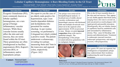
Introduction: Pyogenic Granulomas (PG), more commonly known as lobular capillary hemangiomas, are a rare group of benign inflammatory vascular tumors. While these vascular lesions usually affect the skin and oral mucosa, they are rarely found within the GI tract. These granulomas are often misdiagnosed as bacillary angiomatosis (BA), Kaposi's sarcoma (KS), or inflammatory and/or hyperplastic polyps. Therefore, it is important to report cases of PG so that clinicians also consider it as a differential diagnosis for lower GI bleed.
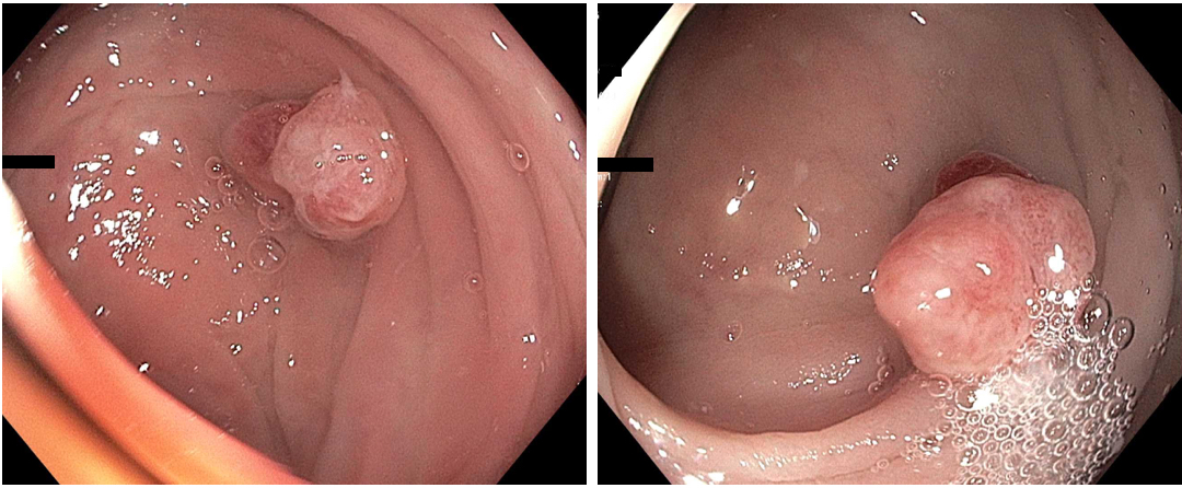
Case Description/Methods: We report to you the case of an elderly male positive for hypertension, type 2 non insulin dependent diabetes and dyslipidemia who presented for routine checkup. As per routine screening, a Cologuard test was done which yielded a positive result. Subsequently, the patient underwent a colonoscopy revealing two polyps measuring 4mm and 7mm in the transverse and sigmoid colon, respectively.
The polyps were resected, retrieved and sent for histopathological analysis. To prevent bleeding, two hemostatic clips were applied, and the area was tattooed with 2mL of spot (carbon black). A localized area of mildly altered vascular, erythematous and hemorrhagic mucosa with thickened folds and multiple diverticula were observed in the sigmoid colon approximately 30-40 cm proximal to anus. Erythema and purulent discharge was also noted at the diverticular opening. Biopsy sample taken from the said area revealed mucosal congestion and reactive epithelial changes. Reports revealed infectious granuloma with no malignant changes.
Discussion: PG lesions of the GI tract exhibit the same histopathological features as those observed in the skin and oral mucosa. They are grossly described as red friable papules that bleed profusely upon minor trauma and range from mm to few cm. Histologically, these present as a cluster of proliferated capillaries in a lobular arrangement lined by a single layer of endothelium with stroma filled with inflammatory cells. Sometimes granulation tissue and neutrophilic infiltrates are also present. These granulomas typically lead to GI bleeding, resulting in anemia and accompanying abdominal pain. Treatment options for PG include endoscopic mucosal resection, endoscopic polypectomy or surgical excision of the granulomas. However, polypectomy is the most commonly utilized modality. Although only a handful cases of gastric PG have been reported in the literature, we believe the incidence is higher as they are frequently mistaken for hyperplastic polyps.

Disclosures:
Hammad Qadri, DO1, Yasir Ahmed, MD1, Nour Gafsi, BSc2, Usama Sakhawat, MD1, Amanke Oranu, MD1. P2007 - Lobular Capillary Hemangioma: A Rare Bleeding Entity in the GI Tract, ACG 2024 Annual Scientific Meeting Abstracts. Philadelphia, PA: American College of Gastroenterology.
1United Health Services, Wilson Medical Center, Binghamton, NY; 2United Health Services, Wilson Medical Center, Johnson City, NY
Introduction: Pyogenic Granulomas (PG), more commonly known as lobular capillary hemangiomas, are a rare group of benign inflammatory vascular tumors. While these vascular lesions usually affect the skin and oral mucosa, they are rarely found within the GI tract. These granulomas are often misdiagnosed as bacillary angiomatosis (BA), Kaposi's sarcoma (KS), or inflammatory and/or hyperplastic polyps. Therefore, it is important to report cases of PG so that clinicians also consider it as a differential diagnosis for lower GI bleed.
Case Description/Methods: We report to you the case of an elderly male positive for hypertension, type 2 non insulin dependent diabetes and dyslipidemia who presented for routine checkup. As per routine screening, a Cologuard test was done which yielded a positive result. Subsequently, the patient underwent a colonoscopy revealing two polyps measuring 4mm and 7mm in the transverse and sigmoid colon, respectively.
The polyps were resected, retrieved and sent for histopathological analysis. To prevent bleeding, two hemostatic clips were applied, and the area was tattooed with 2mL of spot (carbon black). A localized area of mildly altered vascular, erythematous and hemorrhagic mucosa with thickened folds and multiple diverticula were observed in the sigmoid colon approximately 30-40 cm proximal to anus. Erythema and purulent discharge was also noted at the diverticular opening. Biopsy sample taken from the said area revealed mucosal congestion and reactive epithelial changes. Reports revealed infectious granuloma with no malignant changes.
Discussion: PG lesions of the GI tract exhibit the same histopathological features as those observed in the skin and oral mucosa. They are grossly described as red friable papules that bleed profusely upon minor trauma and range from mm to few cm. Histologically, these present as a cluster of proliferated capillaries in a lobular arrangement lined by a single layer of endothelium with stroma filled with inflammatory cells. Sometimes granulation tissue and neutrophilic infiltrates are also present. These granulomas typically lead to GI bleeding, resulting in anemia and accompanying abdominal pain. Treatment options for PG include endoscopic mucosal resection, endoscopic polypectomy or surgical excision of the granulomas. However, polypectomy is the most commonly utilized modality. Although only a handful cases of gastric PG have been reported in the literature, we believe the incidence is higher as they are frequently mistaken for hyperplastic polyps.

Figure: Figure 1 & 2
Disclosures:
Hammad Qadri indicated no relevant financial relationships.
Yasir Ahmed indicated no relevant financial relationships.
Nour Gafsi indicated no relevant financial relationships.
Usama Sakhawat indicated no relevant financial relationships.
Amanke Oranu indicated no relevant financial relationships.
Hammad Qadri, DO1, Yasir Ahmed, MD1, Nour Gafsi, BSc2, Usama Sakhawat, MD1, Amanke Oranu, MD1. P2007 - Lobular Capillary Hemangioma: A Rare Bleeding Entity in the GI Tract, ACG 2024 Annual Scientific Meeting Abstracts. Philadelphia, PA: American College of Gastroenterology.
