Monday Poster Session
Category: Colon
P2014 - Silent Aggressor: Unveiling Duodenal Large-Cell Neuroendocrine Carcinoma
Monday, October 28, 2024
10:30 AM - 4:00 PM ET
Location: Exhibit Hall E

Has Audio

Shyamal Sheth, DO
Franciscan Health Olympia Fields
Chicago, IL
Presenting Author(s)
Shyamal Sheth, DO1, Navin Naik, DO2, Rahil Desai, DO1, Shil Punatar, DO1, Nabiha Haider, DO1, Nick Baur, DO2, Tilemahos Spyratos, DO1
1Franciscan Health Olympia Fields, Chicago, IL; 2Franciscan Health Olympia Fields, Olympia Fields, IL
Introduction: Neuroendocrine tumors (NETs) are rare malignancies, constituting only 0.5% of all cancers, and are predominantly found in females under 50. The majority of NETs occur in the gastrointestinal tract (62-67%) and pulmonary tract (22-27%), with duodenal NETs (D-NETs) making up just 2-3% of gastrointestinal NETs. We present a case of an elderly male who was diagnosed with duodenal large-cell neuroendocrine tumor after an esophagogastroduodenoscopy.
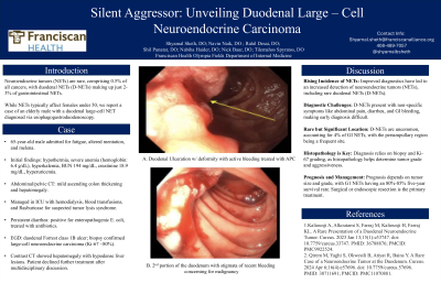
Case Description/Methods: Our patient is a 65-year-old male admitted for fatigue, altered mentation, and melena over the preceding month. On admission, the patient exhibited hypothermia alongside severe anemia (hemoglobin: 6.4 g/dL), hyperkalemia, blood urea nitrogen of 194 mg/dL , serum creatinine of 18.9 mg/dL, and hyperuricemia. Computed tomography (CT) of the abdomen/pelvis revealed mild thickening of the ascending colon up to the hepatic flexure and hepatomegaly. The patient was managed in the intensive care unit (ICU) with emergent hemodialysis due to suspected tumor lysis syndrome, received packed red blood cell transfusions, and rasburicase therapy. Persistent diarrhea prompted an extensive gastrointestinal work-up, revealing positive findings for enteropathogenic Escherichia coli for which antibiotic therapy was initiated. Electrolyte imbalances resolved with hemodialysis, and the patient was transferred to the general medical floor. Given the patient’s profound anemia with transfusion dependence, esophagogastroduodenoscopy (EGD) and colonoscopy were pursued. EGD identified a duodenal Forrest class 1B ulcer concerning for malignancy. Biopsies confirmed a diagnosis of large-cell neuroendocrine carcinoma, with a Ki-67 proliferation index of approximately 80%. Subsequent contrast-enhanced CT of the abdomen/pelvis demonstrated hepatomegaly with numerous hypodense lesions suggestive of malignancy. Following multidisciplinary discussion, the patient opted against further treatment.
Discussion: D-NETs are relatively poorly understood malignancies. Prognosis depends on pathological type, staging, endocrine components, and adjuvant therapy, with lymph node and distant metastases being critical risk factors. Management depends on tumor type, stage, location, size, and histological grade, with resection being the most radical treatment. Given their similarity to other GI tumors and often asymptomatic nature, it is crucial for clinicians to include neuroendocrine tumors in the differential diagnosis for patients with broad symptoms.
Disclosures:
Shyamal Sheth, DO1, Navin Naik, DO2, Rahil Desai, DO1, Shil Punatar, DO1, Nabiha Haider, DO1, Nick Baur, DO2, Tilemahos Spyratos, DO1. P2014 - Silent Aggressor: Unveiling Duodenal Large-Cell Neuroendocrine Carcinoma, ACG 2024 Annual Scientific Meeting Abstracts. Philadelphia, PA: American College of Gastroenterology.
1Franciscan Health Olympia Fields, Chicago, IL; 2Franciscan Health Olympia Fields, Olympia Fields, IL
Introduction: Neuroendocrine tumors (NETs) are rare malignancies, constituting only 0.5% of all cancers, and are predominantly found in females under 50. The majority of NETs occur in the gastrointestinal tract (62-67%) and pulmonary tract (22-27%), with duodenal NETs (D-NETs) making up just 2-3% of gastrointestinal NETs. We present a case of an elderly male who was diagnosed with duodenal large-cell neuroendocrine tumor after an esophagogastroduodenoscopy.
Case Description/Methods: Our patient is a 65-year-old male admitted for fatigue, altered mentation, and melena over the preceding month. On admission, the patient exhibited hypothermia alongside severe anemia (hemoglobin: 6.4 g/dL), hyperkalemia, blood urea nitrogen of 194 mg/dL , serum creatinine of 18.9 mg/dL, and hyperuricemia. Computed tomography (CT) of the abdomen/pelvis revealed mild thickening of the ascending colon up to the hepatic flexure and hepatomegaly. The patient was managed in the intensive care unit (ICU) with emergent hemodialysis due to suspected tumor lysis syndrome, received packed red blood cell transfusions, and rasburicase therapy. Persistent diarrhea prompted an extensive gastrointestinal work-up, revealing positive findings for enteropathogenic Escherichia coli for which antibiotic therapy was initiated. Electrolyte imbalances resolved with hemodialysis, and the patient was transferred to the general medical floor. Given the patient’s profound anemia with transfusion dependence, esophagogastroduodenoscopy (EGD) and colonoscopy were pursued. EGD identified a duodenal Forrest class 1B ulcer concerning for malignancy. Biopsies confirmed a diagnosis of large-cell neuroendocrine carcinoma, with a Ki-67 proliferation index of approximately 80%. Subsequent contrast-enhanced CT of the abdomen/pelvis demonstrated hepatomegaly with numerous hypodense lesions suggestive of malignancy. Following multidisciplinary discussion, the patient opted against further treatment.
Discussion: D-NETs are relatively poorly understood malignancies. Prognosis depends on pathological type, staging, endocrine components, and adjuvant therapy, with lymph node and distant metastases being critical risk factors. Management depends on tumor type, stage, location, size, and histological grade, with resection being the most radical treatment. Given their similarity to other GI tumors and often asymptomatic nature, it is crucial for clinicians to include neuroendocrine tumors in the differential diagnosis for patients with broad symptoms.
Disclosures:
Shyamal Sheth indicated no relevant financial relationships.
Navin Naik indicated no relevant financial relationships.
Rahil Desai indicated no relevant financial relationships.
Shil Punatar indicated no relevant financial relationships.
Nabiha Haider indicated no relevant financial relationships.
Nick Baur indicated no relevant financial relationships.
Tilemahos Spyratos indicated no relevant financial relationships.
Shyamal Sheth, DO1, Navin Naik, DO2, Rahil Desai, DO1, Shil Punatar, DO1, Nabiha Haider, DO1, Nick Baur, DO2, Tilemahos Spyratos, DO1. P2014 - Silent Aggressor: Unveiling Duodenal Large-Cell Neuroendocrine Carcinoma, ACG 2024 Annual Scientific Meeting Abstracts. Philadelphia, PA: American College of Gastroenterology.
