Monday Poster Session
Category: Colon
P2015 - A Rare Case of Colonic Extranodal Marginal Zone Lymphoma Presenting as a Positive FIT Test
Monday, October 28, 2024
10:30 AM - 4:00 PM ET
Location: Exhibit Hall E

Has Audio

Raza Muhammad, DO
University of Chicago Northshore
Chicago, IL
Presenting Author(s)
Raza Muhammad, DO1, Sarah Burroughs, DO2, Mitchelle V. Zolotarevsky, MD3, Robert Toelke, MD4, Matthew Tick, DO5
1University of Chicago Northshore, Evanston, IL; 2University of Chicago, Northshore University Healthsystem, Evanston, IL; 3University of Chicago, NorthShore Internal Medicine, Chicago, IL; 4NorthShore University HealthSystem, Evanston, IL; 5NorthShore University HealthSystem, Highland Park, IL
Introduction: Extranodal Marginal Zone Lymphoma (EMZL) accounts for 5-10% of Non-Hodgkin Lymphoma in the United States. It commonly affects the gastrointestinal tract with the stomach being the most common site but rarely occurs within the colon. We present a rare case of Colonic EMZL diagnosed on screening colonoscopy after a positive fecal immunochemical test (FIT test).
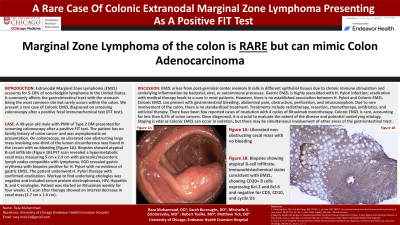
Case Description/Methods: A 49-year-old male with PMH of Type 2 DM presented for screening colonoscopy after a positive FIT test. The patient has no family history of colon cancer and was asymptomatic on presentation. On colonoscopy, an ulcerated non-obstructing large mass involving one-third of the lumen circumference was found in the cecum with no bleeding (Figure 1). Biopsies showed atypical B-cell infiltrate (Figure 2). PET scan revealed a hypermetabolic cecal mass measuring 5 cm x 2.9 cm with paracolic/mesenteric lymph nodes compatible with lymphoma. EGD revealed gastric erythema with biopsies positive for H. Pylori with no evidence of gastric EMZL. The patient underwent H. Pylori therapy with confirmed eradication. Workup to find underlying etiologies was negative and included serum protein electrophoresis, HIV, Hepatitis B, and C serologies. Patient was started on Rituximab weekly for four weeks. CT scan after therapy showed an interval decrease in cecal mass (2.7 cm x 1.6 cm).
Discussion: EMZL arises from post-germinal center memory B cells in different epithelial tissues due to chronic immune stimulation and underlying inflammation by bacterial, viral, or autoimmune processes. Gastric EMZL is highly associated with H. Pylori infection; eradication with medical therapy leads to a cure in most patients. However, there is no established association between H. Pylori and Colonic EMZL. Colonic EMZL can present with gastrointestinal bleeding, abdominal pain, obstruction, perforation, and intussusception. Due to rare involvement of the colon, there is no standardized treatment. Treatments include radiotherapy, resection, chemotherapy, antibiotics, and antiviral therapy. There have been few reported cases of resolution with 4 cycles of Rituximab monotherapy. Colonic EMZL is rare, accounting for less than 0.5% of colon cancers. Once diagnosed, it is crucial to evaluate the extent of the disease and potential underlying etiology. Staging is vital as Colonic EMZL can occur in isolation, but there may be simultaneous involvement of other areas of the gastrointestinal tract.

Disclosures:
Raza Muhammad, DO1, Sarah Burroughs, DO2, Mitchelle V. Zolotarevsky, MD3, Robert Toelke, MD4, Matthew Tick, DO5. P2015 - A Rare Case of Colonic Extranodal Marginal Zone Lymphoma Presenting as a Positive FIT Test, ACG 2024 Annual Scientific Meeting Abstracts. Philadelphia, PA: American College of Gastroenterology.
1University of Chicago Northshore, Evanston, IL; 2University of Chicago, Northshore University Healthsystem, Evanston, IL; 3University of Chicago, NorthShore Internal Medicine, Chicago, IL; 4NorthShore University HealthSystem, Evanston, IL; 5NorthShore University HealthSystem, Highland Park, IL
Introduction: Extranodal Marginal Zone Lymphoma (EMZL) accounts for 5-10% of Non-Hodgkin Lymphoma in the United States. It commonly affects the gastrointestinal tract with the stomach being the most common site but rarely occurs within the colon. We present a rare case of Colonic EMZL diagnosed on screening colonoscopy after a positive fecal immunochemical test (FIT test).
Case Description/Methods: A 49-year-old male with PMH of Type 2 DM presented for screening colonoscopy after a positive FIT test. The patient has no family history of colon cancer and was asymptomatic on presentation. On colonoscopy, an ulcerated non-obstructing large mass involving one-third of the lumen circumference was found in the cecum with no bleeding (Figure 1). Biopsies showed atypical B-cell infiltrate (Figure 2). PET scan revealed a hypermetabolic cecal mass measuring 5 cm x 2.9 cm with paracolic/mesenteric lymph nodes compatible with lymphoma. EGD revealed gastric erythema with biopsies positive for H. Pylori with no evidence of gastric EMZL. The patient underwent H. Pylori therapy with confirmed eradication. Workup to find underlying etiologies was negative and included serum protein electrophoresis, HIV, Hepatitis B, and C serologies. Patient was started on Rituximab weekly for four weeks. CT scan after therapy showed an interval decrease in cecal mass (2.7 cm x 1.6 cm).
Discussion: EMZL arises from post-germinal center memory B cells in different epithelial tissues due to chronic immune stimulation and underlying inflammation by bacterial, viral, or autoimmune processes. Gastric EMZL is highly associated with H. Pylori infection; eradication with medical therapy leads to a cure in most patients. However, there is no established association between H. Pylori and Colonic EMZL. Colonic EMZL can present with gastrointestinal bleeding, abdominal pain, obstruction, perforation, and intussusception. Due to rare involvement of the colon, there is no standardized treatment. Treatments include radiotherapy, resection, chemotherapy, antibiotics, and antiviral therapy. There have been few reported cases of resolution with 4 cycles of Rituximab monotherapy. Colonic EMZL is rare, accounting for less than 0.5% of colon cancers. Once diagnosed, it is crucial to evaluate the extent of the disease and potential underlying etiology. Staging is vital as Colonic EMZL can occur in isolation, but there may be simultaneous involvement of other areas of the gastrointestinal tract.

Figure: (Figure 1): Ulcerated non-obstructing cecal mass with no bleeding
(Figure 2): Biopsies showing atypical B-cell infiltrate. Immunohistochemical stains consistent with EMZL, showing CD20+ B cells expressing Bcl-2 and Bcl-6 and negative for CD5, CD10, and cyclin D1
(Figure 2): Biopsies showing atypical B-cell infiltrate. Immunohistochemical stains consistent with EMZL, showing CD20+ B cells expressing Bcl-2 and Bcl-6 and negative for CD5, CD10, and cyclin D1
Disclosures:
Raza Muhammad indicated no relevant financial relationships.
Sarah Burroughs indicated no relevant financial relationships.
Mitchelle Zolotarevsky indicated no relevant financial relationships.
Robert Toelke indicated no relevant financial relationships.
Matthew Tick indicated no relevant financial relationships.
Raza Muhammad, DO1, Sarah Burroughs, DO2, Mitchelle V. Zolotarevsky, MD3, Robert Toelke, MD4, Matthew Tick, DO5. P2015 - A Rare Case of Colonic Extranodal Marginal Zone Lymphoma Presenting as a Positive FIT Test, ACG 2024 Annual Scientific Meeting Abstracts. Philadelphia, PA: American College of Gastroenterology.
