Monday Poster Session
Category: Biliary/Pancreas
P1783 - Rare Case of Diffuse Large B-cell Lymphoma of the Pancreas
Monday, October 28, 2024
10:30 AM - 4:00 PM ET
Location: Exhibit Hall E

Has Audio
- JS
Jonathan Selzman, DO
UTHSCSA
San Antonio, TX
Presenting Author(s)
Jonathan Selzman, DO1, Dakota Bigham, MD2, James Gnecco, DO1, Hari Sayana, MD3
1UTHSCSA, San Antonio, TX; 2University of Texas Health San Antonio, San Antonio, TX; 3University of Texas Health Science Center, San Antonio, TX
Introduction: While over 90% of pancreatic malignancies present as ductal adenocarcinoma, it is reported that less than 0.5% of all pancreatic neoplasms accounted for are lymphomas. We present a case of a 65-year-old male diagnosed with diffuse large B-cell lymphoma of the pancreas (DLBCL).
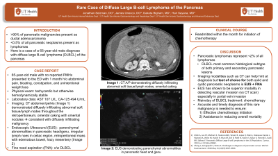
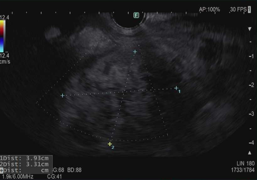
Case Description/Methods: A 65-year-old male with no reported PMHx presented to the emergency department with complaints of abdominal pain, bloating, and unintentional weight loss over the past month. On examination, he was tachycardic but otherwise hemodynamically stable. Physical exam showed a cachectic male with diffuse abdominal tenderness and a positive fluid wave. His labs were significant for an elevation of AST to 107 U/L and CA-19 to 59 U/mL. Computed tomography (CT) of the abdomen and pelvis demonstrated diffusely infiltrating abnormal soft tissue in the retroperitoneum with encasement of aorta, pancreatic body, and spleen with moderate abdominal ascites. Additionally, there was evidence of abnormal lymph nodes throughout the retroperitoneum and omental caking with omental nodules, consistent with diffusely infiltrating malignancy. Advanced endoscopy was consulted, and the patient underwent endoscopic ultrasound (EUS). EUS demonstrated parenchymal abnormalities in the pancreatic head and genu, irregular lymph nodes in the celiac region and a retroperitoneal mass with edematous bowel and hazy mesentery (Figure 1). Pancreas was anatomically identified with endoscope but appeared to have been infiltrated by large retroperitoneal mass. Multiple biopsies were taken, confirming diagnosis of DLBCL. Oncology was consulted, and patient was started on chemotherapy.
Discussion: Pancreatic lymphomas represent < 2% of all lymphomas with DLBCL being the most common histological subtype of both primary and secondary pancreatic lesions. While imaging modalities such as CT can help hint at diagnosis, the gold standard for diagnosis of both solid and cystic pancreatic neoplasms is EUS-FNA to ensure cytopathological certainty. Additionally, EUS has shown to be the superior modality in detecting vascular invasion compared to CT, especially in portal vein invasion. Mainstay of treatment for patients diagnosed with DLBCL of the pancreas is chemotherapy. Overall, accurate and timely diagnosis of this rare malignancy is needed to ensure effective treatment initiation and help to reduce mortality.

Disclosures:
Jonathan Selzman, DO1, Dakota Bigham, MD2, James Gnecco, DO1, Hari Sayana, MD3. P1783 - Rare Case of Diffuse Large B-cell Lymphoma of the Pancreas, ACG 2024 Annual Scientific Meeting Abstracts. Philadelphia, PA: American College of Gastroenterology.
1UTHSCSA, San Antonio, TX; 2University of Texas Health San Antonio, San Antonio, TX; 3University of Texas Health Science Center, San Antonio, TX
Introduction: While over 90% of pancreatic malignancies present as ductal adenocarcinoma, it is reported that less than 0.5% of all pancreatic neoplasms accounted for are lymphomas. We present a case of a 65-year-old male diagnosed with diffuse large B-cell lymphoma of the pancreas (DLBCL).
Case Description/Methods: A 65-year-old male with no reported PMHx presented to the emergency department with complaints of abdominal pain, bloating, and unintentional weight loss over the past month. On examination, he was tachycardic but otherwise hemodynamically stable. Physical exam showed a cachectic male with diffuse abdominal tenderness and a positive fluid wave. His labs were significant for an elevation of AST to 107 U/L and CA-19 to 59 U/mL. Computed tomography (CT) of the abdomen and pelvis demonstrated diffusely infiltrating abnormal soft tissue in the retroperitoneum with encasement of aorta, pancreatic body, and spleen with moderate abdominal ascites. Additionally, there was evidence of abnormal lymph nodes throughout the retroperitoneum and omental caking with omental nodules, consistent with diffusely infiltrating malignancy. Advanced endoscopy was consulted, and the patient underwent endoscopic ultrasound (EUS). EUS demonstrated parenchymal abnormalities in the pancreatic head and genu, irregular lymph nodes in the celiac region and a retroperitoneal mass with edematous bowel and hazy mesentery (Figure 1). Pancreas was anatomically identified with endoscope but appeared to have been infiltrated by large retroperitoneal mass. Multiple biopsies were taken, confirming diagnosis of DLBCL. Oncology was consulted, and patient was started on chemotherapy.
Discussion: Pancreatic lymphomas represent < 2% of all lymphomas with DLBCL being the most common histological subtype of both primary and secondary pancreatic lesions. While imaging modalities such as CT can help hint at diagnosis, the gold standard for diagnosis of both solid and cystic pancreatic neoplasms is EUS-FNA to ensure cytopathological certainty. Additionally, EUS has shown to be the superior modality in detecting vascular invasion compared to CT, especially in portal vein invasion. Mainstay of treatment for patients diagnosed with DLBCL of the pancreas is chemotherapy. Overall, accurate and timely diagnosis of this rare malignancy is needed to ensure effective treatment initiation and help to reduce mortality.

Figure: Figure 1. Endoscopic ultrasound images of irregular mass in the retroperitoneum.
Disclosures:
Jonathan Selzman indicated no relevant financial relationships.
Dakota Bigham indicated no relevant financial relationships.
James Gnecco indicated no relevant financial relationships.
Hari Sayana indicated no relevant financial relationships.
Jonathan Selzman, DO1, Dakota Bigham, MD2, James Gnecco, DO1, Hari Sayana, MD3. P1783 - Rare Case of Diffuse Large B-cell Lymphoma of the Pancreas, ACG 2024 Annual Scientific Meeting Abstracts. Philadelphia, PA: American College of Gastroenterology.
