Sunday Poster Session
Category: Stomach
P1706 - Gastric Mass Mystery: Revealing Extracavitary Primary Effusion Lymphoma
Sunday, October 27, 2024
3:30 PM - 7:00 PM ET
Location: Exhibit Hall E

Has Audio

Iqra Kazi, MD
University of Texas Health San Antonio
San Antonio, TX
Presenting Author(s)
Iqra Kazi, MD, Saatchi Kuwelker, MD, Juan Echavarria, MD, Apeksha Agarwal, MD
University of Texas Health San Antonio, San Antonio, TX
Introduction: Primary effusion lymphoma (PEL) is a rare, aggressive B-cell lymphoma, manifesting as malignant effusions in body cavities without discrete solid masses. However, extracavitary solid variants can develop, often in the peritoneum, pleural space and pericardium. We present a rare case found in the gastric mucosa.
Case Description/Methods: 51-year-old male with HIV/AIDS (viral load 1150, CD4 count 47) was admitted to the ICU due to undifferentiated shock and severe anemia (hemoglobin (Hgb) 4.9). During a complicated hospital course (including bacteremia and respiratory failure) he was found to have nodular thickening of the stomach wall measuring 2.7cm on CT abdomen. He had no GI symptoms. Laboratory studies were significant for pancytopenia (15,000) and normocytic anemia (MCV 91). Upper endoscopy showed a superficial esophageal ulcer and an ulcerated, infiltrated mass-like appearance of the mucosa extended from gastric cardia to the proximal antrum. Pathology revealed atypical cells with plasmablastic morphology expressing MUM1, partial CD138, CD30, EBER-ISH, HHV8, and lacked CD20, consistent with diagnosis of extracavitary PEL. He later developed hemorrhagic shock necessitating blood transfusions with presumed bleeding from PEL. He was deemed a poor candidate for rituximab due to low CD4 count and high risk of opportunistic infections. He decided to pursue comfort care and passed away.
Discussion: PEL predominantly affects elderly or immunocompromised individuals particularly those diagnosed with HIV infection or post-solid organ transplantation. PEL is also associated with human herpesvirus 8 (Kaposi Sarcoma-associated herpesvirus). This malignancy accounts for 4% of all HIV-associated non-Hodgkin Lymphoma, male-predominant, with median diagnosis age 44-45 years. Clinical manifestations mainly present with effusions in the body cavity. A variant of PEL may lead to development of lymphoma in lymph nodes or as a solid tumor mass in extranodal sites (GI tract, lung, skin or central nervous system).
We share this case of PEL presenting as a stomach mass with likely occult GI bleeding. Only one other case has been reported in the literature. Extracavitary variants of PEL are very rare and diagnostically challenging as in this case without GI symptoms. Immunocompromised patients with imaging irregularities should be thoroughly evaluated through endoscopy with biopsies to rule out rare pathology such as this which may be lifesaving with early intervention.

Disclosures:
Iqra Kazi, MD, Saatchi Kuwelker, MD, Juan Echavarria, MD, Apeksha Agarwal, MD. P1706 - Gastric Mass Mystery: Revealing Extracavitary Primary Effusion Lymphoma, ACG 2024 Annual Scientific Meeting Abstracts. Philadelphia, PA: American College of Gastroenterology.
University of Texas Health San Antonio, San Antonio, TX
Introduction: Primary effusion lymphoma (PEL) is a rare, aggressive B-cell lymphoma, manifesting as malignant effusions in body cavities without discrete solid masses. However, extracavitary solid variants can develop, often in the peritoneum, pleural space and pericardium. We present a rare case found in the gastric mucosa.
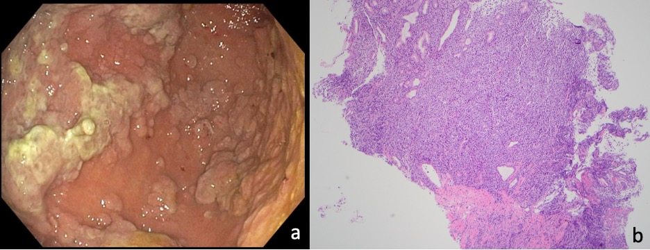
Case Description/Methods: 51-year-old male with HIV/AIDS (viral load 1150, CD4 count 47) was admitted to the ICU due to undifferentiated shock and severe anemia (hemoglobin (Hgb) 4.9). During a complicated hospital course (including bacteremia and respiratory failure) he was found to have nodular thickening of the stomach wall measuring 2.7cm on CT abdomen. He had no GI symptoms. Laboratory studies were significant for pancytopenia (15,000) and normocytic anemia (MCV 91). Upper endoscopy showed a superficial esophageal ulcer and an ulcerated, infiltrated mass-like appearance of the mucosa extended from gastric cardia to the proximal antrum. Pathology revealed atypical cells with plasmablastic morphology expressing MUM1, partial CD138, CD30, EBER-ISH, HHV8, and lacked CD20, consistent with diagnosis of extracavitary PEL. He later developed hemorrhagic shock necessitating blood transfusions with presumed bleeding from PEL. He was deemed a poor candidate for rituximab due to low CD4 count and high risk of opportunistic infections. He decided to pursue comfort care and passed away.
Discussion: PEL predominantly affects elderly or immunocompromised individuals particularly those diagnosed with HIV infection or post-solid organ transplantation. PEL is also associated with human herpesvirus 8 (Kaposi Sarcoma-associated herpesvirus). This malignancy accounts for 4% of all HIV-associated non-Hodgkin Lymphoma, male-predominant, with median diagnosis age 44-45 years. Clinical manifestations mainly present with effusions in the body cavity. A variant of PEL may lead to development of lymphoma in lymph nodes or as a solid tumor mass in extranodal sites (GI tract, lung, skin or central nervous system).
We share this case of PEL presenting as a stomach mass with likely occult GI bleeding. Only one other case has been reported in the literature. Extracavitary variants of PEL are very rare and diagnostically challenging as in this case without GI symptoms. Immunocompromised patients with imaging irregularities should be thoroughly evaluated through endoscopy with biopsies to rule out rare pathology such as this which may be lifesaving with early intervention.

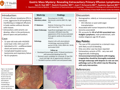
Figure: Figure 1 (a) ulcerated and infiltrated appearance of gastric mucosa seen on EGD found to have (b) atypical cells with plasmablastic morphology on histopathology concerning for primary effusion lymphoma.
Disclosures:
Iqra Kazi indicated no relevant financial relationships.
Saatchi Kuwelker indicated no relevant financial relationships.
Juan Echavarria indicated no relevant financial relationships.
Apeksha Agarwal indicated no relevant financial relationships.
Iqra Kazi, MD, Saatchi Kuwelker, MD, Juan Echavarria, MD, Apeksha Agarwal, MD. P1706 - Gastric Mass Mystery: Revealing Extracavitary Primary Effusion Lymphoma, ACG 2024 Annual Scientific Meeting Abstracts. Philadelphia, PA: American College of Gastroenterology.
