Sunday Poster Session
Category: Stomach
P1707 - Gastric Granulomas: An Unusual Presentation of Sarcoidosis
Sunday, October 27, 2024
3:30 PM - 7:00 PM ET
Location: Exhibit Hall E

Has Audio
- AJ
Alexander Jenkins, DO
National Capital Consortium
Bethesda, MD
Presenting Author(s)
Alexander Jenkins, DO1, Roy K. Wong, MD, MACG2
1National Capital Consortium, Bethesda, MD; 2Walter Reed National Military Medical Center, Bethesda, MD
Introduction: Sarcoidosis of the GI tract is rare but can affect tissue from the esophagus to the rectum. We present a case of a Hispanic male presenting with anemia, early satiety, unexplained 60 lb. weight loss, and a diagnoses of GI sarcoidosis.
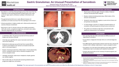
Case Description/Methods: The patient is a 39 yr. old healthy male presenting with 3 weeks of fatigue, shortness of breath and severe vitamin B12 deficiency anemia. EGD revealed diffuse erythema and friability of the stomach and duodenal bulb with pyloric narrowing. Colonoscopy was normal including the terminal ileum. Biopsies revealed granulomatous inflammation in the duodenal bulb, antrum, and gastric body as well as intestinal metaplasia in the antrum and gastric body. Histologic staining for autoimmune metaplastic atrophic gastritis was negative. Nine months later, he presented with worsening abdominal pain, progressive 60 lb. weight loss, and iron deficiency anemia. EGD showed nodular thickening of the stomach and duodenal bulb with biopsies revealing findings similar to previous histology. Histologic staining showed no evidence of tuberculosis (TB), fungal elements, foreign bodies, eosinophils, granulomatous arteritis, syphilis, or lymphoma. Serologic evaluation was negative for TB, anti-intrinsic factor antibodies, and anti-parietal cell antibodies. Cross sectional imaging was obtained with incidental pulmonary findings highly suggestive of sarcoidosis. The patient was initiated on high-dose corticosteroids with significant improvement in his symptoms.
Discussion: This case illustrates the rarity and difficulty in diagnosing GI sarcoidosis. The differential for granulomatous diseases of the GI tract is broad and includes Crohn's disease, TB, H. pylori, and gastric cancer. We believe this patient's iron and vitamin B12 deficiency is related to malabsorption due to sarcoidosis with duodenal involvement. The compelling radiographic evidence of pulmonary sarcoidosis along with the absence of key features seen in Crohn’s disease or autoimmune atrophic gastritis and a negative infectious workup makes gastric and duodenal sarcoidosis the most likely unifying diagnosis.

Disclosures:
Alexander Jenkins, DO1, Roy K. Wong, MD, MACG2. P1707 - Gastric Granulomas: An Unusual Presentation of Sarcoidosis, ACG 2024 Annual Scientific Meeting Abstracts. Philadelphia, PA: American College of Gastroenterology.
1National Capital Consortium, Bethesda, MD; 2Walter Reed National Military Medical Center, Bethesda, MD
Introduction: Sarcoidosis of the GI tract is rare but can affect tissue from the esophagus to the rectum. We present a case of a Hispanic male presenting with anemia, early satiety, unexplained 60 lb. weight loss, and a diagnoses of GI sarcoidosis.
Case Description/Methods: The patient is a 39 yr. old healthy male presenting with 3 weeks of fatigue, shortness of breath and severe vitamin B12 deficiency anemia. EGD revealed diffuse erythema and friability of the stomach and duodenal bulb with pyloric narrowing. Colonoscopy was normal including the terminal ileum. Biopsies revealed granulomatous inflammation in the duodenal bulb, antrum, and gastric body as well as intestinal metaplasia in the antrum and gastric body. Histologic staining for autoimmune metaplastic atrophic gastritis was negative. Nine months later, he presented with worsening abdominal pain, progressive 60 lb. weight loss, and iron deficiency anemia. EGD showed nodular thickening of the stomach and duodenal bulb with biopsies revealing findings similar to previous histology. Histologic staining showed no evidence of tuberculosis (TB), fungal elements, foreign bodies, eosinophils, granulomatous arteritis, syphilis, or lymphoma. Serologic evaluation was negative for TB, anti-intrinsic factor antibodies, and anti-parietal cell antibodies. Cross sectional imaging was obtained with incidental pulmonary findings highly suggestive of sarcoidosis. The patient was initiated on high-dose corticosteroids with significant improvement in his symptoms.
Discussion: This case illustrates the rarity and difficulty in diagnosing GI sarcoidosis. The differential for granulomatous diseases of the GI tract is broad and includes Crohn's disease, TB, H. pylori, and gastric cancer. We believe this patient's iron and vitamin B12 deficiency is related to malabsorption due to sarcoidosis with duodenal involvement. The compelling radiographic evidence of pulmonary sarcoidosis along with the absence of key features seen in Crohn’s disease or autoimmune atrophic gastritis and a negative infectious workup makes gastric and duodenal sarcoidosis the most likely unifying diagnosis.

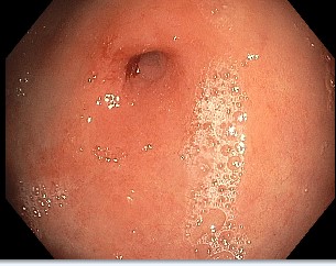
Figure: Antral nodularity with stenosed pylorus
Disclosures:
Alexander Jenkins indicated no relevant financial relationships.
Roy Wong indicated no relevant financial relationships.
Alexander Jenkins, DO1, Roy K. Wong, MD, MACG2. P1707 - Gastric Granulomas: An Unusual Presentation of Sarcoidosis, ACG 2024 Annual Scientific Meeting Abstracts. Philadelphia, PA: American College of Gastroenterology.

