Sunday Poster Session
Category: Stomach
P1632 - Ocular Metastases: A Rare Presentation of Gastric Adenocarcinoma
Sunday, October 27, 2024
3:30 PM - 7:00 PM ET
Location: Exhibit Hall E

Has Audio
- SP
Susie Park, MD
George Washington University Hospital
Washington, DC
Presenting Author(s)
Susie Park, MD1, Alisa Malyavko, MS2, Adam Jacob, MD2, Giancarlo Colon Rosa, MD2, Marie L.. Borum, MD, MPH, FACG2, Samuel A.. Schueler, MD2
1George Washington University Hospital, Washington, DC; 2George Washington University School of Medicine and Health Sciences, Washington, DC
Introduction: Gastric adenocarcinoma with distant metastases caries an estimated 5-year survival of 7%. Ocular metastases are rare, usually identified after the primary tumor, and associated with poor prognosis. We present a patient with ocular symptoms prior to identification of gastric adenocarcinoma.
Case Description/Methods: A 53-year-old male with hypertension presented with abdominal pain, nausea, vomiting, and melena. He reported two months of double vision, swelling, and throbbing of the right eye. He was evaluated by ophthalmology and treated for presumed orbital cellulitis with antibiotics followed by prednisone due to non-response.
On admission, patient was hypertensive. Labs showed normocytic anemia (Hemoglobin 10 gm/dL) elevated liver enzymes (Aspartate transaminase 126 units/L, Alanine aminotransferase 104 units/L, Alkaline phosphatase 4.3units/L) and elevated lipase (519 units/L). Facial exam showed proptosis, eyelid edema, and conjunctival injection of the right eye. Abdominal exam showed distention and diffuse tenderness.
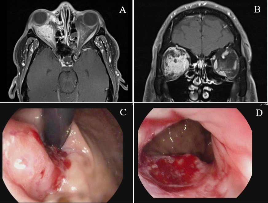
Magnetic resonance imaging (MRI) of the head and neck showed right orbital mass extending from the eyelid to the orbital apex involving the medial and inferior rectus, and surrounding the optic nerve (Figure 1). Computed tomography of the abdomen showed distal gastric wall thickening, gastrohepatic ligament adenopathy, mesenteric adenopathy, nodularity of the omentum concerning for peritoneal carcinomatosis, liver hypodensities and indeterminate 1.0-centimeter lesion within the T11 vertebral body.
Esophagogastroduodenoscopy (EGD) identified erythematous, friable, nodular and ulcerated mucosa in the gastroesophageal junction and gastric cardia (Figure 1). Biopsies revealed gastric adenocarcinoma with mucinous component. Ocular biopsy showed poorly differentiated adenocarcinoma with mucinous features.
Discussion: Ocular metastases of gastric cancer (GC) are rare. They most often occur in the choroid followed by the iris, eyelid, conjunctiva and optic nerve. A survey of 520 patients with GC found choroidal metastasis in only 4% of patients. A study analyzing case reports from 2011 - 2019 reported only five cases of choroidal metastases from GC. Another study reviewing twelve cases of metastatic GC to the eyelid found eyelid metastases as initial presentation in only four cases. This case highlights the need to consider metastases as a possible cause for ocular symptoms and recognize this rare manifestation of GC.

Disclosures:
Susie Park, MD1, Alisa Malyavko, MS2, Adam Jacob, MD2, Giancarlo Colon Rosa, MD2, Marie L.. Borum, MD, MPH, FACG2, Samuel A.. Schueler, MD2. P1632 - Ocular Metastases: A Rare Presentation of Gastric Adenocarcinoma, ACG 2024 Annual Scientific Meeting Abstracts. Philadelphia, PA: American College of Gastroenterology.
1George Washington University Hospital, Washington, DC; 2George Washington University School of Medicine and Health Sciences, Washington, DC
Introduction: Gastric adenocarcinoma with distant metastases caries an estimated 5-year survival of 7%. Ocular metastases are rare, usually identified after the primary tumor, and associated with poor prognosis. We present a patient with ocular symptoms prior to identification of gastric adenocarcinoma.
Case Description/Methods: A 53-year-old male with hypertension presented with abdominal pain, nausea, vomiting, and melena. He reported two months of double vision, swelling, and throbbing of the right eye. He was evaluated by ophthalmology and treated for presumed orbital cellulitis with antibiotics followed by prednisone due to non-response.
On admission, patient was hypertensive. Labs showed normocytic anemia (Hemoglobin 10 gm/dL) elevated liver enzymes (Aspartate transaminase 126 units/L, Alanine aminotransferase 104 units/L, Alkaline phosphatase 4.3units/L) and elevated lipase (519 units/L). Facial exam showed proptosis, eyelid edema, and conjunctival injection of the right eye. Abdominal exam showed distention and diffuse tenderness.
Magnetic resonance imaging (MRI) of the head and neck showed right orbital mass extending from the eyelid to the orbital apex involving the medial and inferior rectus, and surrounding the optic nerve (Figure 1). Computed tomography of the abdomen showed distal gastric wall thickening, gastrohepatic ligament adenopathy, mesenteric adenopathy, nodularity of the omentum concerning for peritoneal carcinomatosis, liver hypodensities and indeterminate 1.0-centimeter lesion within the T11 vertebral body.
Esophagogastroduodenoscopy (EGD) identified erythematous, friable, nodular and ulcerated mucosa in the gastroesophageal junction and gastric cardia (Figure 1). Biopsies revealed gastric adenocarcinoma with mucinous component. Ocular biopsy showed poorly differentiated adenocarcinoma with mucinous features.
Discussion: Ocular metastases of gastric cancer (GC) are rare. They most often occur in the choroid followed by the iris, eyelid, conjunctiva and optic nerve. A survey of 520 patients with GC found choroidal metastasis in only 4% of patients. A study analyzing case reports from 2011 - 2019 reported only five cases of choroidal metastases from GC. Another study reviewing twelve cases of metastatic GC to the eyelid found eyelid metastases as initial presentation in only four cases. This case highlights the need to consider metastases as a possible cause for ocular symptoms and recognize this rare manifestation of GC.

Figure: Figure 1. Panels A and B represent MRI images of the face, neck and orbit showing right orbital mass (A) Horizontal section and (B) Coronal section. Panels C and D represent EGD images of the gastric mass (C) Gastroesophageal junction in retroflexion and (D) gastric cardia
Disclosures:
Susie Park indicated no relevant financial relationships.
Alisa Malyavko indicated no relevant financial relationships.
Adam Jacob indicated no relevant financial relationships.
Giancarlo Colon Rosa indicated no relevant financial relationships.
Marie Borum: Takeda Pharmaceuticals – Consultant, Speakers Bureau.
Samuel Schueler indicated no relevant financial relationships.
Susie Park, MD1, Alisa Malyavko, MS2, Adam Jacob, MD2, Giancarlo Colon Rosa, MD2, Marie L.. Borum, MD, MPH, FACG2, Samuel A.. Schueler, MD2. P1632 - Ocular Metastases: A Rare Presentation of Gastric Adenocarcinoma, ACG 2024 Annual Scientific Meeting Abstracts. Philadelphia, PA: American College of Gastroenterology.
