Sunday Poster Session
Category: Liver
P1359 - Hepatic Sarcoidosis: A Rare Symptomatic Case of Isolated Hepatic Sarcoidosis
Sunday, October 27, 2024
3:30 PM - 7:00 PM ET
Location: Exhibit Hall E

Has Audio
- ST
Shunsa Tarar, DO
Albany Medical Center
Albany, NY
Presenting Author(s)
Shunsa Tarar, DO, Elie Tannous, MD, Dimple Ghassi, MD
Albany Medical Center, Albany, NY
Introduction: Sarcoidosis is a systemic disease characterized by non-caseating granulomas in affected organs, most commonly the lungs. It has a prevalence of about 4.7 in 100,000 and an incidence of 1.0–35.5 in 100,000 per year. The highest rates are reported in northern European and African-American women, with the lowest in Japan. Most patients with hepatic sarcoidosis are asymptomatic and don't require treatment. We present a rare case of symptomatic hepatic sarcoidosis with abnormal transaminases.
Case Description/Methods: The patient is a 67-year-old Asian female who presented with symptoms of weight loss, pruritus, and bloating for six months. Medical history includes diabetes, hypothyroidism, hyperlipidemia, and osteoporosis. She takes Glipizide, Levothyroxine, Lisinopril, Metformin, Risedronate, and Rosuvastatin.
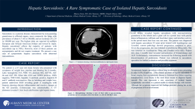
Laboratory findings were notable for AST 66 and ALP 198. Due to symptoms of belching and weight loss the patient underwent an EGD, which revealed grade A esophagitis with biopsies showing chronic inflammation. Colonoscopy was unremarkable. A CT abdomen revealed a 1.3 cm focal calcification in the right hepatic dome.
A liver biopsy revealed hepatic sarcoidosis with non-necrotizing granuloma in the lobule and a giant cell in a portal tract. The patient had been diagnosed with hepatic sarcoidosis 22 years prior and was treated with Azathioprine and Ursodiol. Current pathology showed progression compared to prior findings.
Given the patient's progression and symptoms, she was initiated on prednisone 40mg daily. However, steroids were tapered within a week due to a spike in blood sugar levels. She was restarted on Azathioprine 50mg and Ursodiol twice daily. Repeat labs three months later revealed normalization of transaminases and ALP. Unfortunately, transaminases increased the following month with the discontinuation of prednisone. The patient was referred to a sarcoidosis specialist for further evaluation and possible treatment.
Discussion: Symptomatic hepatic sarcoidosis without lung involvement is rare, presenting in only 5-15% of patients. The clinical spectrum of hepatic sarcoidosis is broad, ranging from asymptomatic disease to fulminant liver failure requiring transplantation. Liver biopsy is the most direct means to diagnose hepatic sarcoid. This case represents a rare occurrence of isolated symptomatic hepatic sarcoidosis in a patient unable to tolerate therapy with steroids. Although her symptoms improved, lab findings persisted, placing her at risk of progression of liver involvement.

Disclosures:
Shunsa Tarar, DO, Elie Tannous, MD, Dimple Ghassi, MD. P1359 - Hepatic Sarcoidosis: A Rare Symptomatic Case of Isolated Hepatic Sarcoidosis, ACG 2024 Annual Scientific Meeting Abstracts. Philadelphia, PA: American College of Gastroenterology.
Albany Medical Center, Albany, NY
Introduction: Sarcoidosis is a systemic disease characterized by non-caseating granulomas in affected organs, most commonly the lungs. It has a prevalence of about 4.7 in 100,000 and an incidence of 1.0–35.5 in 100,000 per year. The highest rates are reported in northern European and African-American women, with the lowest in Japan. Most patients with hepatic sarcoidosis are asymptomatic and don't require treatment. We present a rare case of symptomatic hepatic sarcoidosis with abnormal transaminases.
Case Description/Methods: The patient is a 67-year-old Asian female who presented with symptoms of weight loss, pruritus, and bloating for six months. Medical history includes diabetes, hypothyroidism, hyperlipidemia, and osteoporosis. She takes Glipizide, Levothyroxine, Lisinopril, Metformin, Risedronate, and Rosuvastatin.
Laboratory findings were notable for AST 66 and ALP 198. Due to symptoms of belching and weight loss the patient underwent an EGD, which revealed grade A esophagitis with biopsies showing chronic inflammation. Colonoscopy was unremarkable. A CT abdomen revealed a 1.3 cm focal calcification in the right hepatic dome.
A liver biopsy revealed hepatic sarcoidosis with non-necrotizing granuloma in the lobule and a giant cell in a portal tract. The patient had been diagnosed with hepatic sarcoidosis 22 years prior and was treated with Azathioprine and Ursodiol. Current pathology showed progression compared to prior findings.
Given the patient's progression and symptoms, she was initiated on prednisone 40mg daily. However, steroids were tapered within a week due to a spike in blood sugar levels. She was restarted on Azathioprine 50mg and Ursodiol twice daily. Repeat labs three months later revealed normalization of transaminases and ALP. Unfortunately, transaminases increased the following month with the discontinuation of prednisone. The patient was referred to a sarcoidosis specialist for further evaluation and possible treatment.
Discussion: Symptomatic hepatic sarcoidosis without lung involvement is rare, presenting in only 5-15% of patients. The clinical spectrum of hepatic sarcoidosis is broad, ranging from asymptomatic disease to fulminant liver failure requiring transplantation. Liver biopsy is the most direct means to diagnose hepatic sarcoid. This case represents a rare occurrence of isolated symptomatic hepatic sarcoidosis in a patient unable to tolerate therapy with steroids. Although her symptoms improved, lab findings persisted, placing her at risk of progression of liver involvement.

Figure: Figure 1. CT image showing a 1.3 cm focal calcification in the right hepatic dome (A). Histopathology showing a core of liver with a non-necrotizing granuloma (arrow) and giant cells in the lobule (B: x10; C: X20 H&E stain).
Disclosures:
Shunsa Tarar indicated no relevant financial relationships.
Elie Tannous indicated no relevant financial relationships.
Dimple Ghassi indicated no relevant financial relationships.
Shunsa Tarar, DO, Elie Tannous, MD, Dimple Ghassi, MD. P1359 - Hepatic Sarcoidosis: A Rare Symptomatic Case of Isolated Hepatic Sarcoidosis, ACG 2024 Annual Scientific Meeting Abstracts. Philadelphia, PA: American College of Gastroenterology.
