Sunday Poster Session
Category: Liver
P1284 - A Challenging Diagnosis of a Liver Mass: Inflammatory Pseudotumor of the Liver
Sunday, October 27, 2024
3:30 PM - 7:00 PM ET
Location: Exhibit Hall E

Has Audio

Bianca Thakkar, DO
University of Connecticut Health Center
Farmington, CT
Presenting Author(s)
Bianca Thakkar, DO1, Anum Dileep, MBBS2, Marianna Scranton, DO2
1University of Connecticut Health Center, Farmington, CT; 2Hartford Hospital, Hartford, CT
Introduction: Inflammatory pseudotumor (IPT) is a rare benign lesion described as chronic infiltration of inflammatory cells with fibrosis. While IPTs most commonly occur in the lungs, IPT of the liver (IPTL) has an incidence of 0.7%. The etiology and pathogenesis of IPTL is unknown. Here we present a case of IPTL masquerading as a liver abscess.
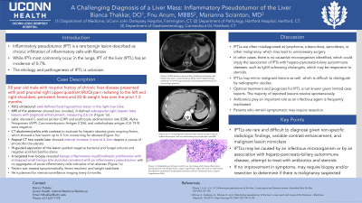
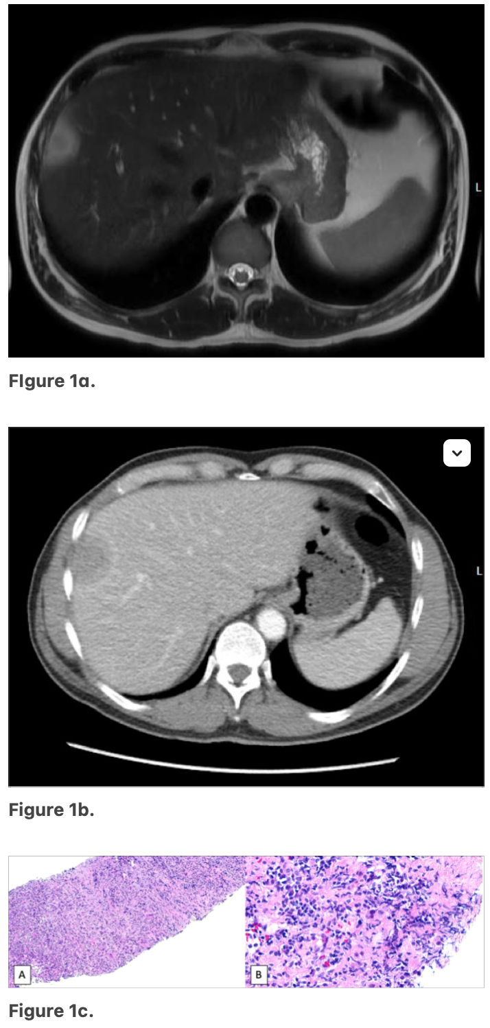
Case Description/Methods: A 58 year old male with no prior history of chronic liver disease presented with post prandial right upper quadrant (RUQ) pain and radiating to the left and right shoulders, persistent fevers and 20-lb weight loss over the prior 1.5 months. RUQ ultrasound showed a normal gallbladder and biliary tract with a well-defined focal hypoechoic lesion in the right liver lobe. MRI of the abdomen showed two rounded, ill-defined subcapsular right hepatic lobe lesions with targetoid enhancement, measuring 2.6 cm (Figure 1a). Labs were unremarkable except for elevated C-reactive protein (CRP) and erythrocyte sedimentation rate (ESR). Alpha Fetoprotein (AFP), Carcinoembryonic Antigen (CEA), and carbohydrate antigen (CA 19-9) were negative. His QuantiFERON-TB gold returned positive, but he had a negative chest x-ray. He underwent CT of the abdomen and pelvis with contrast to evaluate for hepatic abscess given ongoing fevers, which showed a liver lesion up to 3.1cm concerning for abscess (Figure 1b). Blood cultures were negative. A repeat CT two weeks later showed interval increase in size to 4.3cm despite a course of amoxicillin/clavulanate. IR-guided aspiration of the lesion yielded negative bacterial and fungal cultures and negative acid-fast bacillus stains. A targeted liver biopsy revealed benign inflammatory myofibroblastic proliferation with entrapped small benign bile ductules consistent with an inflammatory pseudotumor with no aggregates of acute inflammatory cells indicative of an abscess (Figure 1c). Patient was treated symptomatically, fevers resolve, and weight stabilized. He is planned for interval surveillance imaging in 6 months.
Discussion: IPTLs pose a diagnostic challenge given their nonspecific laboratory and imaging findings. They are often misdiagnosed as lymphoma, tuberculosis, sarcoidosis, or other malignancy, which may lead to unnecessary surgery. Optimal treatment and prognosis for IPTL is not known given limited case reports. The majority of reported lesions resolve spontaneously. Some patients improve with antibiotics or corticosteroids alone. Patients who remain symptomatic may require resection.

Disclosures:
Bianca Thakkar, DO1, Anum Dileep, MBBS2, Marianna Scranton, DO2. P1284 - A Challenging Diagnosis of a Liver Mass: Inflammatory Pseudotumor of the Liver, ACG 2024 Annual Scientific Meeting Abstracts. Philadelphia, PA: American College of Gastroenterology.
1University of Connecticut Health Center, Farmington, CT; 2Hartford Hospital, Hartford, CT
Introduction: Inflammatory pseudotumor (IPT) is a rare benign lesion described as chronic infiltration of inflammatory cells with fibrosis. While IPTs most commonly occur in the lungs, IPT of the liver (IPTL) has an incidence of 0.7%. The etiology and pathogenesis of IPTL is unknown. Here we present a case of IPTL masquerading as a liver abscess.
Case Description/Methods: A 58 year old male with no prior history of chronic liver disease presented with post prandial right upper quadrant (RUQ) pain and radiating to the left and right shoulders, persistent fevers and 20-lb weight loss over the prior 1.5 months. RUQ ultrasound showed a normal gallbladder and biliary tract with a well-defined focal hypoechoic lesion in the right liver lobe. MRI of the abdomen showed two rounded, ill-defined subcapsular right hepatic lobe lesions with targetoid enhancement, measuring 2.6 cm (Figure 1a). Labs were unremarkable except for elevated C-reactive protein (CRP) and erythrocyte sedimentation rate (ESR). Alpha Fetoprotein (AFP), Carcinoembryonic Antigen (CEA), and carbohydrate antigen (CA 19-9) were negative. His QuantiFERON-TB gold returned positive, but he had a negative chest x-ray. He underwent CT of the abdomen and pelvis with contrast to evaluate for hepatic abscess given ongoing fevers, which showed a liver lesion up to 3.1cm concerning for abscess (Figure 1b). Blood cultures were negative. A repeat CT two weeks later showed interval increase in size to 4.3cm despite a course of amoxicillin/clavulanate. IR-guided aspiration of the lesion yielded negative bacterial and fungal cultures and negative acid-fast bacillus stains. A targeted liver biopsy revealed benign inflammatory myofibroblastic proliferation with entrapped small benign bile ductules consistent with an inflammatory pseudotumor with no aggregates of acute inflammatory cells indicative of an abscess (Figure 1c). Patient was treated symptomatically, fevers resolve, and weight stabilized. He is planned for interval surveillance imaging in 6 months.
Discussion: IPTLs pose a diagnostic challenge given their nonspecific laboratory and imaging findings. They are often misdiagnosed as lymphoma, tuberculosis, sarcoidosis, or other malignancy, which may lead to unnecessary surgery. Optimal treatment and prognosis for IPTL is not known given limited case reports. The majority of reported lesions resolve spontaneously. Some patients improve with antibiotics or corticosteroids alone. Patients who remain symptomatic may require resection.

Figure: Figure 1a. MRI abdomen demonstrating ill-defined subcapsular right hepatic lobe lesion, characterized by diffuse mild T2 hyperintensity, peripheral rim-enhancing component, central hypoenhancement, and surrounding hyperemia.
Figure 1b. CT of the abdomen and pelvis with interval increase in size of hepatic dome lesion, with rim enhancement one month after initial MRI.
Figure 1c. Histopathologic findings, A and B Liver core biopsy which shows inflammatory pseudotumor (A. Hematoxylin-Eosin, original magnification 100x), with a 0.5 cm inflammatory pseudotumor composed of lymphocytes and plasma cells (B. Hematoxylin-Eosin, original magnification 200x).
Disclosures:
Bianca Thakkar indicated no relevant financial relationships.
Anum Dileep indicated no relevant financial relationships.
Marianna Scranton indicated no relevant financial relationships.
Bianca Thakkar, DO1, Anum Dileep, MBBS2, Marianna Scranton, DO2. P1284 - A Challenging Diagnosis of a Liver Mass: Inflammatory Pseudotumor of the Liver, ACG 2024 Annual Scientific Meeting Abstracts. Philadelphia, PA: American College of Gastroenterology.
