Sunday Poster Session
Category: Interventional Endoscopy
P1082 - Endoscopic Ultrasound-Directed Trans-Enteric ERCP (EDEE) in a Patient With Malignant Afferent Limb and Hepaticojejunostomy Stricture
Sunday, October 27, 2024
3:30 PM - 7:00 PM ET
Location: Exhibit Hall E

Has Audio

Layth Alzubaidy, MD
University of Texas Health Science Center
Tyler, TX
Presenting Author(s)
Award: Presidential Poster Award
Layth Alzubaidy, MD1, Muhammad Almas, MD2, Muhammad Baig, MD3
1University of Texas Health Science Center, Tyler, TX; 2University of Texas Health Sciences Center, Tyler, TX; 3UT Health Science Center, Tyler, TX
Introduction: Lumen-apposing metal stents (LAMS) are being used more frequently in patients with surgically altered anatomy, offering a less invasive alternative to procedures like percutaneous or surgical interventions.
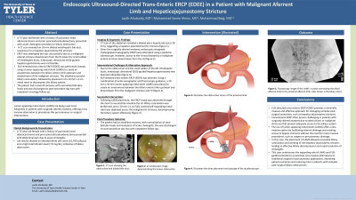
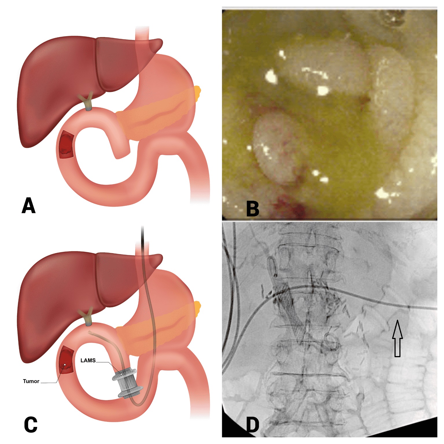
Case Description/Methods: A 72-year-old female with a history of pancreatic head adenocarcinoma and pancreaticoduodenectomy presented to our hospital with abdominal pain secondary to acute cholangitis. She was hemodynamically stable, but laboratory evaluation revealed an elevated white cell count of 16,700 cells per microliter and a total bilirubin level of 7.9 mg/dl. A computed tomography scan of the abdomen showed post-surgical anatomy with a dilated extra-hepatic bile duct to 18 mm suspicious of hepatico-jejunostomy (HJ) stricture. Endoscopic retrograde cholangiopancreatography (ERCP) was attempted using a pediatric colonoscope, but HJ was not accessible due to a malignant enteral stricture just downstream to the HJ (figure 1A and 1B). Endoscopic ultrasound-guided hepatico-gastrostomy was not feasible due to the small caliber of left intra-hepatic ducts. Therefore, EUS-directed trans-enteric ERCP (EDEE) was planned. Using endo-sonographic and fluoroscopic guidance a 20 mm x 10 mm lumen-apposing metal stent (LAMS) was used to create an anastomosis between the biliary enteric limb upstream and downstream from the malignant stricture and HJ (Figure 1C and 1D). Consequently, an ERCP scope could be advanced through the LAMS to visualize HJ. Biliary cannulation was successful and a 10 mm x 4 cm fully covered self-expanding metal stent was placed across malignant HJ stricture to successfully decompress the biliary system. The patient had an excellent post-procedure course with normalization of total bilirubin and resolution of acute cholangitis. She was discharged on post-procedure day two with outpatient oncology follow-up.
Discussion: This case highlights a novel approach of EUS-directed trans-enteric ERCP (EDEE) in managing patients with complex post-surgical anatomies.

Disclosures:
Layth Alzubaidy, MD1, Muhammad Almas, MD2, Muhammad Baig, MD3. P1082 - Endoscopic Ultrasound-Directed Trans-Enteric ERCP (EDEE) in a Patient With Malignant Afferent Limb and Hepaticojejunostomy Stricture, ACG 2024 Annual Scientific Meeting Abstracts. Philadelphia, PA: American College of Gastroenterology.
Layth Alzubaidy, MD1, Muhammad Almas, MD2, Muhammad Baig, MD3
1University of Texas Health Science Center, Tyler, TX; 2University of Texas Health Sciences Center, Tyler, TX; 3UT Health Science Center, Tyler, TX
Introduction: Lumen-apposing metal stents (LAMS) are being used more frequently in patients with surgically altered anatomy, offering a less invasive alternative to procedures like percutaneous or surgical interventions.
Case Description/Methods: A 72-year-old female with a history of pancreatic head adenocarcinoma and pancreaticoduodenectomy presented to our hospital with abdominal pain secondary to acute cholangitis. She was hemodynamically stable, but laboratory evaluation revealed an elevated white cell count of 16,700 cells per microliter and a total bilirubin level of 7.9 mg/dl. A computed tomography scan of the abdomen showed post-surgical anatomy with a dilated extra-hepatic bile duct to 18 mm suspicious of hepatico-jejunostomy (HJ) stricture. Endoscopic retrograde cholangiopancreatography (ERCP) was attempted using a pediatric colonoscope, but HJ was not accessible due to a malignant enteral stricture just downstream to the HJ (figure 1A and 1B). Endoscopic ultrasound-guided hepatico-gastrostomy was not feasible due to the small caliber of left intra-hepatic ducts. Therefore, EUS-directed trans-enteric ERCP (EDEE) was planned. Using endo-sonographic and fluoroscopic guidance a 20 mm x 10 mm lumen-apposing metal stent (LAMS) was used to create an anastomosis between the biliary enteric limb upstream and downstream from the malignant stricture and HJ (Figure 1C and 1D). Consequently, an ERCP scope could be advanced through the LAMS to visualize HJ. Biliary cannulation was successful and a 10 mm x 4 cm fully covered self-expanding metal stent was placed across malignant HJ stricture to successfully decompress the biliary system. The patient had an excellent post-procedure course with normalization of total bilirubin and resolution of acute cholangitis. She was discharged on post-procedure day two with outpatient oncology follow-up.
Discussion: This case highlights a novel approach of EUS-directed trans-enteric ERCP (EDEE) in managing patients with complex post-surgical anatomies.

Figure: Figure 1: (A) illustrates the obstructive tumor of the proximal limb. (B) an endoscopic image demonstrating the lumen obstruction by the tumor. (C) illustrates the LAMS placement and passage of the duodenoscope. (D) fluoroscopy image of the LAMS (arrow) in place connecting the distal afferent limb to the proximal afferent limb. Also shown is the biliary stent.
Disclosures:
Layth Alzubaidy indicated no relevant financial relationships.
Muhammad Almas indicated no relevant financial relationships.
Muhammad Baig indicated no relevant financial relationships.
Layth Alzubaidy, MD1, Muhammad Almas, MD2, Muhammad Baig, MD3. P1082 - Endoscopic Ultrasound-Directed Trans-Enteric ERCP (EDEE) in a Patient With Malignant Afferent Limb and Hepaticojejunostomy Stricture, ACG 2024 Annual Scientific Meeting Abstracts. Philadelphia, PA: American College of Gastroenterology.

