Sunday Poster Session
Category: GI Bleeding
P0755 - A Rare Case of Isolated Jejunal Varices in the Absence of Portal Hypertension
Sunday, October 27, 2024
3:30 PM - 7:00 PM ET
Location: Exhibit Hall E

Has Audio

Hannah Darnell, DO
University of Kentucky
Lexington, KY
Presenting Author(s)
Hannah Darnell, DO, Aaron Brenner, MD, Marijeta Pekez, MD
University of Kentucky, Lexington, KY
Introduction: Ectopic varices are rare portosystemic collaterals located outside the gastroesophageal region that cause only about 5% of GI bleeding. Specifically, small intestinal varices are even rarer and pose unique diagnostic and therapeutic challenges. We present a rare case of a 19-year-old male with hematochezia discovered to be secondary to jejunal varices in the absence of portal hypertension.
Case Description/Methods: A 19-year-old male with no past medical history presented to the emergency department with one episode of hematochezia and subsequent syncope. Labs revealed hemoglobin 13.9 g/dL and the remainder labs were normal. His hemoglobin dropped to 6.3 g/dL within 48 hours.
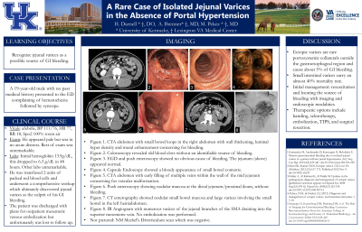
His workup included extensive imaging starting with computed tomography angiography (CTA) of his abdomen and pelvis that revealed small bowel luminal hyperdensity and mural enhancement during the venous phase concerning for bleeding. EGD and push enteroscopy revealed no obvious cause of bleeding. Colonoscopy revealed old blood clots within the entire colon without an identifiable source of bleeding. Capsule endoscopy showed blood 50% through the small bowel without a visualized lesion. Repeat CTA revealed early filling of a dilated vein associated with mid jejunal loops concerning for an underlying vascular malformation. Meckles scan was negative. CT enterography (CTE) was remarkable for nodular small bowel mucosa with large varices involving the small bowel. IR guided angiogram found numerous varices, most pronounced in the left midabdominal jejunal branches, that drained into the superior mesenteric vein. No embolization was performed. The patient was scheduled to follow up with IR in two weeks, however he was lost to follow up.
Discussion: Ectopic varices commonly occur secondary to portal hypertension; however, they can develop in its absence. Among ectopic varices, small intestinal varices are rare and carry an almost 40% mortality rate. Initial management is focused on resuscitation and locating the source of bleeding with imaging and endoscopic modalities. EGD and colonoscopy should be performed but if inconclusive, imaging for suspected small bowel bleeding includes CTA, CTE, capsule endoscopy, and Meckles scan. Therapeutic options include banding, sclerotherapy, embolization, TIPS, and surgical resection. Although ectopic varices are rare, given their high mortality, it’s important for physicians to consider this diagnosis in a patient with GI bleeding when upper and lower endoscopy are inconclusive.
Disclosures:
Hannah Darnell, DO, Aaron Brenner, MD, Marijeta Pekez, MD. P0755 - A Rare Case of Isolated Jejunal Varices in the Absence of Portal Hypertension, ACG 2024 Annual Scientific Meeting Abstracts. Philadelphia, PA: American College of Gastroenterology.
University of Kentucky, Lexington, KY
Introduction: Ectopic varices are rare portosystemic collaterals located outside the gastroesophageal region that cause only about 5% of GI bleeding. Specifically, small intestinal varices are even rarer and pose unique diagnostic and therapeutic challenges. We present a rare case of a 19-year-old male with hematochezia discovered to be secondary to jejunal varices in the absence of portal hypertension.
Case Description/Methods: A 19-year-old male with no past medical history presented to the emergency department with one episode of hematochezia and subsequent syncope. Labs revealed hemoglobin 13.9 g/dL and the remainder labs were normal. His hemoglobin dropped to 6.3 g/dL within 48 hours.
His workup included extensive imaging starting with computed tomography angiography (CTA) of his abdomen and pelvis that revealed small bowel luminal hyperdensity and mural enhancement during the venous phase concerning for bleeding. EGD and push enteroscopy revealed no obvious cause of bleeding. Colonoscopy revealed old blood clots within the entire colon without an identifiable source of bleeding. Capsule endoscopy showed blood 50% through the small bowel without a visualized lesion. Repeat CTA revealed early filling of a dilated vein associated with mid jejunal loops concerning for an underlying vascular malformation. Meckles scan was negative. CT enterography (CTE) was remarkable for nodular small bowel mucosa with large varices involving the small bowel. IR guided angiogram found numerous varices, most pronounced in the left midabdominal jejunal branches, that drained into the superior mesenteric vein. No embolization was performed. The patient was scheduled to follow up with IR in two weeks, however he was lost to follow up.
Discussion: Ectopic varices commonly occur secondary to portal hypertension; however, they can develop in its absence. Among ectopic varices, small intestinal varices are rare and carry an almost 40% mortality rate. Initial management is focused on resuscitation and locating the source of bleeding with imaging and endoscopic modalities. EGD and colonoscopy should be performed but if inconclusive, imaging for suspected small bowel bleeding includes CTA, CTE, capsule endoscopy, and Meckles scan. Therapeutic options include banding, sclerotherapy, embolization, TIPS, and surgical resection. Although ectopic varices are rare, given their high mortality, it’s important for physicians to consider this diagnosis in a patient with GI bleeding when upper and lower endoscopy are inconclusive.
Disclosures:
Hannah Darnell indicated no relevant financial relationships.
Aaron Brenner indicated no relevant financial relationships.
Marijeta Pekez indicated no relevant financial relationships.
Hannah Darnell, DO, Aaron Brenner, MD, Marijeta Pekez, MD. P0755 - A Rare Case of Isolated Jejunal Varices in the Absence of Portal Hypertension, ACG 2024 Annual Scientific Meeting Abstracts. Philadelphia, PA: American College of Gastroenterology.
