Sunday Poster Session
Category: Esophagus
P0608 - Cerebral Air Embolism Due to Atrio-Esophageal Fistula: A Case Report and Literature Review
Sunday, October 27, 2024
3:30 PM - 7:00 PM ET
Location: Exhibit Hall E

Has Audio
- YS
Yazan Sallam, MD
University of Missouri - Kansas City School of Medicine
Kansas City, MO
Presenting Author(s)
Yazan Sallam, MD1, Adel Muhanna, MD1, Rajiv Chhabra, MD1, Mohammad Adam, MD2
1University of Missouri - Kansas City School of Medicine, Kansas City, MO; 2University of Missouri, Kansas City, MO
Introduction: Cerebral air embolism is a rare but serious complication, usually caused by trauma or an iatrogenic event. A rare cause of cerebral air embolism is an Atrio-esophageal fistula (AEF), a fatal complication that usually happens after atrial fibrillation (AFib) ablation. We describe here a patient who presented to our institution with altered mental status and was unfortunate to have an AEF that was complicated by cerebral air embolism leading to his death.
Case Description/Methods: A 65-year-old male patient who was admitted to our hospital with 1 day history of altered mental status. Patient is known case of AFib that was recently treated at an outside facility with radiofrequency ablation 3 weeks prior to admission. On presentation, patient had a Glasco coma scale score of 9. Brain Magnetic resonance imaging (MRI) showed acute multiple infarcts involving his right cerebral hemisphere, suggestive of an embolic pathology.
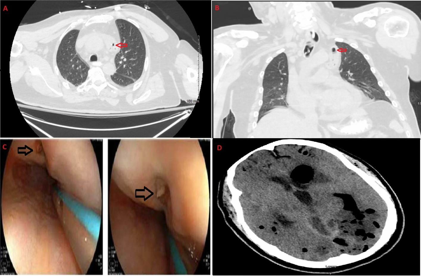
Patient was intubated and admitted to the Intensive care unit (ICU). CT scan of the chest with oral contrast showed a foci of air in the mid-distal esophagus but without an extravasation of contrast, moderate pericardial effusion with fluid collection containing foci of air in the superior mediastinum(figure A,B). Patient underwent a midline sternotomy, with findings of purulent discharge, along with a posterior left atrial perforation near the left inferior pulmonary vein, the perforation was repaired, and drains were inserted. At this point, Cardiothoracic surgeons requested to perform an endoscopy to confirm the AEF, giving that the myocardium was perforated but the pericardium was sealed.
Esophagogastroduodenoscopy (EGD) was done, showed an anterior deep mid-esophageal defect, around 8-millimeter wide, which appeared to be sealed off on inspection(figure C). Patient later went into cardiac arrest and was resuscitated. CT head was done and showed extensive gas throughout the brain(figure D). Nuclear imaging showed no intracerebral flow and confirmed brain death.
Discussion: AEF is a rare but often fatal complication that happens usually after catheter-ablation for AF. Our patient had an ablation done 3 weeks prior to presentation and developed an AEF, which ended with an air embolism. Diagnosis of AEF is usually done using CT or MRI of the chest with water-soluble oral contrast. Endoscopy should usually be avoided due to air insufflation. Although there are no guidelines for the treatment of AEF, recent studies lean towards surgical management over endoscopic stent treatment.

Disclosures:
Yazan Sallam, MD1, Adel Muhanna, MD1, Rajiv Chhabra, MD1, Mohammad Adam, MD2. P0608 - Cerebral Air Embolism Due to Atrio-Esophageal Fistula: A Case Report and Literature Review, ACG 2024 Annual Scientific Meeting Abstracts. Philadelphia, PA: American College of Gastroenterology.
1University of Missouri - Kansas City School of Medicine, Kansas City, MO; 2University of Missouri, Kansas City, MO
Introduction: Cerebral air embolism is a rare but serious complication, usually caused by trauma or an iatrogenic event. A rare cause of cerebral air embolism is an Atrio-esophageal fistula (AEF), a fatal complication that usually happens after atrial fibrillation (AFib) ablation. We describe here a patient who presented to our institution with altered mental status and was unfortunate to have an AEF that was complicated by cerebral air embolism leading to his death.
Case Description/Methods: A 65-year-old male patient who was admitted to our hospital with 1 day history of altered mental status. Patient is known case of AFib that was recently treated at an outside facility with radiofrequency ablation 3 weeks prior to admission. On presentation, patient had a Glasco coma scale score of 9. Brain Magnetic resonance imaging (MRI) showed acute multiple infarcts involving his right cerebral hemisphere, suggestive of an embolic pathology.
Patient was intubated and admitted to the Intensive care unit (ICU). CT scan of the chest with oral contrast showed a foci of air in the mid-distal esophagus but without an extravasation of contrast, moderate pericardial effusion with fluid collection containing foci of air in the superior mediastinum(figure A,B). Patient underwent a midline sternotomy, with findings of purulent discharge, along with a posterior left atrial perforation near the left inferior pulmonary vein, the perforation was repaired, and drains were inserted. At this point, Cardiothoracic surgeons requested to perform an endoscopy to confirm the AEF, giving that the myocardium was perforated but the pericardium was sealed.
Esophagogastroduodenoscopy (EGD) was done, showed an anterior deep mid-esophageal defect, around 8-millimeter wide, which appeared to be sealed off on inspection(figure C). Patient later went into cardiac arrest and was resuscitated. CT head was done and showed extensive gas throughout the brain(figure D). Nuclear imaging showed no intracerebral flow and confirmed brain death.
Discussion: AEF is a rare but often fatal complication that happens usually after catheter-ablation for AF. Our patient had an ablation done 3 weeks prior to presentation and developed an AEF, which ended with an air embolism. Diagnosis of AEF is usually done using CT or MRI of the chest with water-soluble oral contrast. Endoscopy should usually be avoided due to air insufflation. Although there are no guidelines for the treatment of AEF, recent studies lean towards surgical management over endoscopic stent treatment.

Figure: Figure A and B. Foci of air in the Superior mediastinum.
Figure C. Anterior deep mid-esophageal defect.
Figure D. Multiple Cerebral Air embolus.
Figure C. Anterior deep mid-esophageal defect.
Figure D. Multiple Cerebral Air embolus.
Disclosures:
Yazan Sallam indicated no relevant financial relationships.
Adel Muhanna indicated no relevant financial relationships.
Rajiv Chhabra indicated no relevant financial relationships.
Mohammad Adam indicated no relevant financial relationships.
Yazan Sallam, MD1, Adel Muhanna, MD1, Rajiv Chhabra, MD1, Mohammad Adam, MD2. P0608 - Cerebral Air Embolism Due to Atrio-Esophageal Fistula: A Case Report and Literature Review, ACG 2024 Annual Scientific Meeting Abstracts. Philadelphia, PA: American College of Gastroenterology.
