Sunday Poster Session
Category: Esophagus
P0549 - Navigating Dysphagia: A Rare Case of Granular Cell Tumor
Sunday, October 27, 2024
3:30 PM - 7:00 PM ET
Location: Exhibit Hall E

Has Audio

Feruza Abraamyan, MD
Sutter Health
Roseville, CA
Presenting Author(s)
Feruza Abraamyan, MD, Lingala Shilpa, MD
Sutter Health, Roseville, CA
Introduction: Knowing rare etiologies of dysphagia and determining appropriate diagnostic, treatment, and surveillance workups are essential in gastroenterology practice. Here, we present a rare case of a symptomatic esophageal mass.
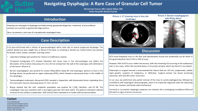
Case Description/Methods: A 61-year-old female with a history of gastroesophageal reflux came due to severe progressive dysphagia. The patient denied any pain, weight loss, or blood in the stool, no smoking or alcohol use. Family history was positive for breast, colon, ovarian, prostate, and lung cancer. Laboratory findings were positive for chronic iron deficiency anemia. Computed tomography (CT) showed lobulated soft tissue mass in the mid-esophagus just before the bifurcation of the trachea measuring 19 x 16 x 20 mm arising from the wall of the esophagus with obliteration of the lumen. Barium esophagogram was positive for convex filling defect along the mid esophagus. Due to the findings above, we performed an upper endoscopy (EGD), which showed a submucosal lesion in the middle of the esophagus. Transesophageal endoscopic ultrasound (EUS) revealed a hypoechoic well-demarcated lesion originating from the muscularis mucosa measuring about 17 mm x 16 mm. Biopsy showed that the cells' neoplastic population was positive for S-100, Calretinin, and CD 68. The esophageal mass was consistent with a neurogenic granular cell tumor (GCT). The patient underwent subtotal removal of mass without complication. EGD and EUS were repeated in 6 months without abnormalities.
Discussion: The majority of gastrointestinal stromal tumors (GCTs) are found in the skin and subcutaneous tissues, but in 6% to 10% of cases, they can be located in the gastrointestinal tract. However, 95% of GCTs are within the mucosa, with the remaining 5% in the submucosa and, in rare cases, within the neuronal plexus in muscularis propria, as was the case with our patient. It is recommended to remove lesions that are >10 mm, symptomatic, exhibit rapid growth, have suspicion of malignancy, or infiltration. Surgical excision has shown promising outcomes, with only 8% tumor recurrence. In our case, we performed partial removal of the mass to avoid esophagectomy, followed by surveillance with esophageal ultrasonography. Currently, our patient's dysphagia symptoms have resolved, and she is undergoing surveillance EGD and EUS with no signs of tumor recurrence.
Disclosures:
Feruza Abraamyan, MD, Lingala Shilpa, MD. P0549 - Navigating Dysphagia: A Rare Case of Granular Cell Tumor, ACG 2024 Annual Scientific Meeting Abstracts. Philadelphia, PA: American College of Gastroenterology.
Sutter Health, Roseville, CA
Introduction: Knowing rare etiologies of dysphagia and determining appropriate diagnostic, treatment, and surveillance workups are essential in gastroenterology practice. Here, we present a rare case of a symptomatic esophageal mass.
Case Description/Methods: A 61-year-old female with a history of gastroesophageal reflux came due to severe progressive dysphagia. The patient denied any pain, weight loss, or blood in the stool, no smoking or alcohol use. Family history was positive for breast, colon, ovarian, prostate, and lung cancer. Laboratory findings were positive for chronic iron deficiency anemia. Computed tomography (CT) showed lobulated soft tissue mass in the mid-esophagus just before the bifurcation of the trachea measuring 19 x 16 x 20 mm arising from the wall of the esophagus with obliteration of the lumen. Barium esophagogram was positive for convex filling defect along the mid esophagus. Due to the findings above, we performed an upper endoscopy (EGD), which showed a submucosal lesion in the middle of the esophagus. Transesophageal endoscopic ultrasound (EUS) revealed a hypoechoic well-demarcated lesion originating from the muscularis mucosa measuring about 17 mm x 16 mm. Biopsy showed that the cells' neoplastic population was positive for S-100, Calretinin, and CD 68. The esophageal mass was consistent with a neurogenic granular cell tumor (GCT). The patient underwent subtotal removal of mass without complication. EGD and EUS were repeated in 6 months without abnormalities.
Discussion: The majority of gastrointestinal stromal tumors (GCTs) are found in the skin and subcutaneous tissues, but in 6% to 10% of cases, they can be located in the gastrointestinal tract. However, 95% of GCTs are within the mucosa, with the remaining 5% in the submucosa and, in rare cases, within the neuronal plexus in muscularis propria, as was the case with our patient. It is recommended to remove lesions that are >10 mm, symptomatic, exhibit rapid growth, have suspicion of malignancy, or infiltration. Surgical excision has shown promising outcomes, with only 8% tumor recurrence. In our case, we performed partial removal of the mass to avoid esophagectomy, followed by surveillance with esophageal ultrasonography. Currently, our patient's dysphagia symptoms have resolved, and she is undergoing surveillance EGD and EUS with no signs of tumor recurrence.
Disclosures:
Feruza Abraamyan indicated no relevant financial relationships.
Lingala Shilpa indicated no relevant financial relationships.
Feruza Abraamyan, MD, Lingala Shilpa, MD. P0549 - Navigating Dysphagia: A Rare Case of Granular Cell Tumor, ACG 2024 Annual Scientific Meeting Abstracts. Philadelphia, PA: American College of Gastroenterology.
