Sunday Poster Session
Category: Esophagus
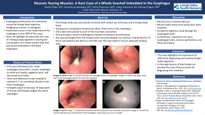
P0556 - Mussels Tearing Muscles: A Rare Case of a Whole Seashell Imbedded in the Esophagus
Sunday, October 27, 2024
3:30 PM - 7:00 PM ET
Location: Exhibit Hall E

Has Audio
.jpg)
Rachel Patel, DO
Lehigh Valley Health Network
Coopersburg, PA
Presenting Author(s)
Rachel Patel, DO1, Amanda Jacubowsky, DO2, Romy Chamoun, MD2, Gracy Chamoun, BS3, Michael Engels, MD4
1Lehigh Valley Health Network, Coopersburg, PA; 2Lehigh Valley Health Network, Allentown, PA; 3University of Balamand, Arlington, VA; 4Eastern Pennsylvania Gastrointestinal and Liver Specialists, Allentown, PA
Introduction: Esophageal perforations are commonly caused by foreign body ingestion, malignancy, trauma, or iatrogenic. Perforations occur in the distal third of the esophagus in over 90% of the case due to weaker musculature in the dorsolateral esophageal wall. Here, we highlight an extremely rare case of a foreign body ingestion involving the consumption of a whole mussel shell that was found embedded in the distal esophagus.
Case Description/Methods: A 65-year-old female with no pertinent past medical history presented to the hospital with lower esophageal discomfort. She was on vacation in Puerto Rico six days before arrival and endorsed sudden onset nausea, dysphagia to solids and liquids, epigastric pain, and decreased oral intake. She was seen by a medical clinic in Puerto Rico, in which x-rays revealed a retained foreign body in the esophagus with no signs of perforation. She was informed that the object would pass, and no intervention was recommended. Upon arrival to the United States, she presented to the emergency department for further evaluation. Lab tests were within normal limitations. Repeat chest and abdominal x-rays revealed a 3.7 cm curvilinear density in the lower esophagus. Due to increased risk of esophageal perforation, the patient underwent an emergent upper endoscopy. She was found to have a large piece of a mussel shell deeply lodged into the contralateral walls of the distal esophagus. The foreign body was successfully removed with careful use of forceps and a foreign body hood. Subsequent visualization revealed two deep, linear tears in the esophagus where the shell was impacted. Dura-clips were placed to each of the traumatic ulcerations. Post-procedure, the patient underwent a barium esophagram which showed no contrast extravasation or evidence of perforation. She was discharged from the hospital with recommendations to continue a liquid diet for 72 hours and advance her diet to a soft diet over the next week to ensure adequate healing time.
Discussion: Mussels are a common delicacy cooked around the globe. It is known that mussel shells break more easily than other seashells. Accidental ingestion could damage the esophageal walls. Furthermore, impaction can cause esophageal tears, erosion, perforation, and fistula formation. This case highlights the importance of effectively diagnosing and treating foreign body ingestions. A thorough history of food intake can provide the most clinical acumen for diagnosing this condition.

Disclosures:
Rachel Patel, DO1, Amanda Jacubowsky, DO2, Romy Chamoun, MD2, Gracy Chamoun, BS3, Michael Engels, MD4. P0556 - Mussels Tearing Muscles: A Rare Case of a Whole Seashell Imbedded in the Esophagus, ACG 2024 Annual Scientific Meeting Abstracts. Philadelphia, PA: American College of Gastroenterology.
1Lehigh Valley Health Network, Coopersburg, PA; 2Lehigh Valley Health Network, Allentown, PA; 3University of Balamand, Arlington, VA; 4Eastern Pennsylvania Gastrointestinal and Liver Specialists, Allentown, PA
Introduction: Esophageal perforations are commonly caused by foreign body ingestion, malignancy, trauma, or iatrogenic. Perforations occur in the distal third of the esophagus in over 90% of the case due to weaker musculature in the dorsolateral esophageal wall. Here, we highlight an extremely rare case of a foreign body ingestion involving the consumption of a whole mussel shell that was found embedded in the distal esophagus.
Case Description/Methods: A 65-year-old female with no pertinent past medical history presented to the hospital with lower esophageal discomfort. She was on vacation in Puerto Rico six days before arrival and endorsed sudden onset nausea, dysphagia to solids and liquids, epigastric pain, and decreased oral intake. She was seen by a medical clinic in Puerto Rico, in which x-rays revealed a retained foreign body in the esophagus with no signs of perforation. She was informed that the object would pass, and no intervention was recommended. Upon arrival to the United States, she presented to the emergency department for further evaluation. Lab tests were within normal limitations. Repeat chest and abdominal x-rays revealed a 3.7 cm curvilinear density in the lower esophagus. Due to increased risk of esophageal perforation, the patient underwent an emergent upper endoscopy. She was found to have a large piece of a mussel shell deeply lodged into the contralateral walls of the distal esophagus. The foreign body was successfully removed with careful use of forceps and a foreign body hood. Subsequent visualization revealed two deep, linear tears in the esophagus where the shell was impacted. Dura-clips were placed to each of the traumatic ulcerations. Post-procedure, the patient underwent a barium esophagram which showed no contrast extravasation or evidence of perforation. She was discharged from the hospital with recommendations to continue a liquid diet for 72 hours and advance her diet to a soft diet over the next week to ensure adequate healing time.
Discussion: Mussels are a common delicacy cooked around the globe. It is known that mussel shells break more easily than other seashells. Accidental ingestion could damage the esophageal walls. Furthermore, impaction can cause esophageal tears, erosion, perforation, and fistula formation. This case highlights the importance of effectively diagnosing and treating foreign body ingestions. A thorough history of food intake can provide the most clinical acumen for diagnosing this condition.

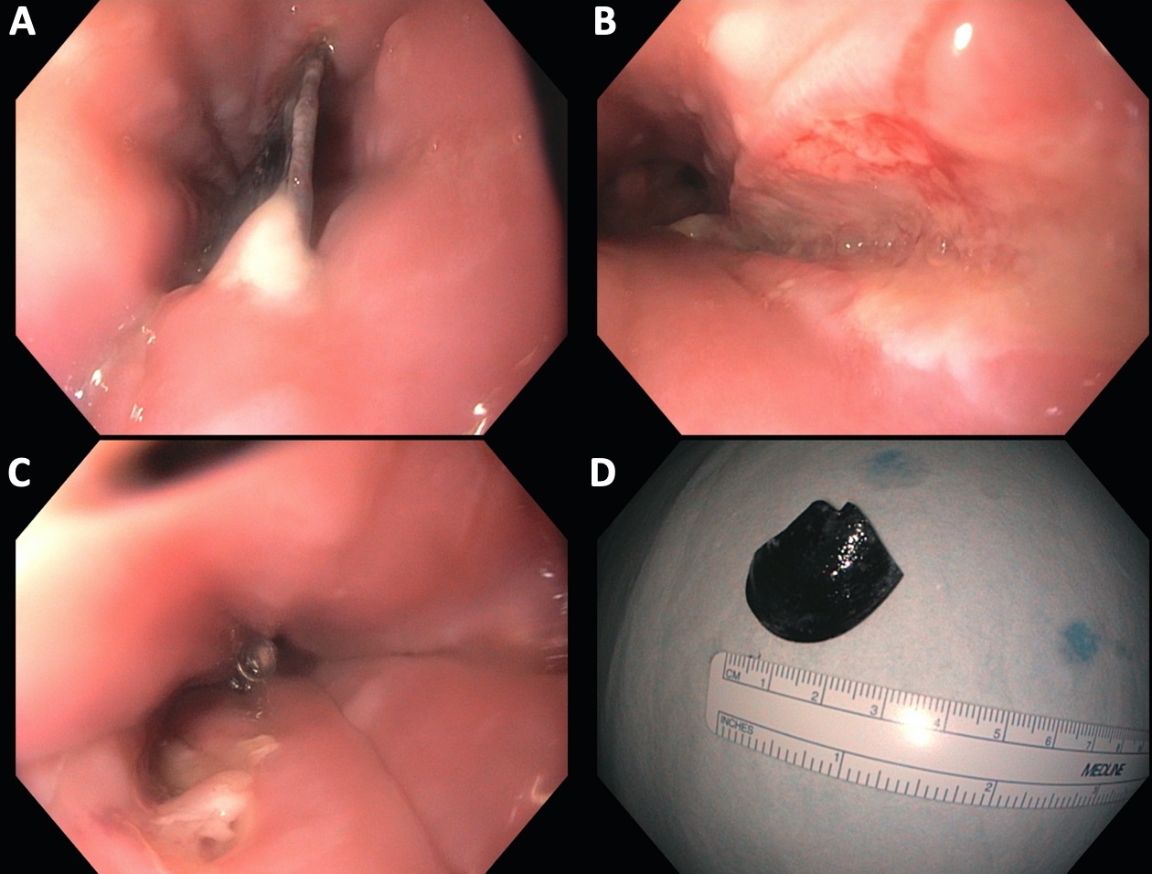
Figure: Image 'A' reveals a mussel shell dislodged into the esophagus. Image 'B' reveals two deep, linear tears in the esophagus where the shell was impacted. Image 'C' reveals the DuraClips that were placed to the traumatic ulcerations. Image 'D' reveals the mussel shell that was retrieved from the esophagus, measuring up to 3 cm in length.
Disclosures:
Rachel Patel indicated no relevant financial relationships.
Amanda Jacubowsky indicated no relevant financial relationships.
Romy Chamoun indicated no relevant financial relationships.
Gracy Chamoun indicated no relevant financial relationships.
Michael Engels indicated no relevant financial relationships.
Rachel Patel, DO1, Amanda Jacubowsky, DO2, Romy Chamoun, MD2, Gracy Chamoun, BS3, Michael Engels, MD4. P0556 - Mussels Tearing Muscles: A Rare Case of a Whole Seashell Imbedded in the Esophagus, ACG 2024 Annual Scientific Meeting Abstracts. Philadelphia, PA: American College of Gastroenterology.
