Sunday Poster Session
Category: Esophagus
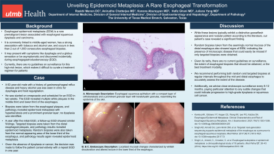
P0560 - Unveiling Epidermoid Metaplasia: A Rare Esophageal Transformation
Sunday, October 27, 2024
3:30 PM - 7:00 PM ET
Location: Exhibit Hall E

Has Audio
- RM
Raakhi Menon, DO
University of Texas Medical Branch
Galveston, TX
Presenting Author(s)
Raakhi Menon, DO, Anirudha Chatterjee, MD, Kanana Aburayyan, MD, Hafiz Ghani, MD, Juliana Yang, MD
University of Texas Medical Branch, Galveston, TX
Introduction: Esophageal epidermoid metaplasia (EEM) is a rare premalignant lesion associated with esophageal squamous dysplasia and carcinoma. It is commonly linked to middle-aged women, has a strong association with tobacco and alcohol use, and occurs in less than 2 out of 1,000 consecutive esophageal biopsies. It may present with symptoms like dysphagia and a globus sensation or be asymptomatic and discovered incidentally during esophagogastroduodenoscopy (EGD). Currently, there are no guidelines on surveillance for this high-risk lesion, which makes it difficult to curate a treatment regimen for patients.
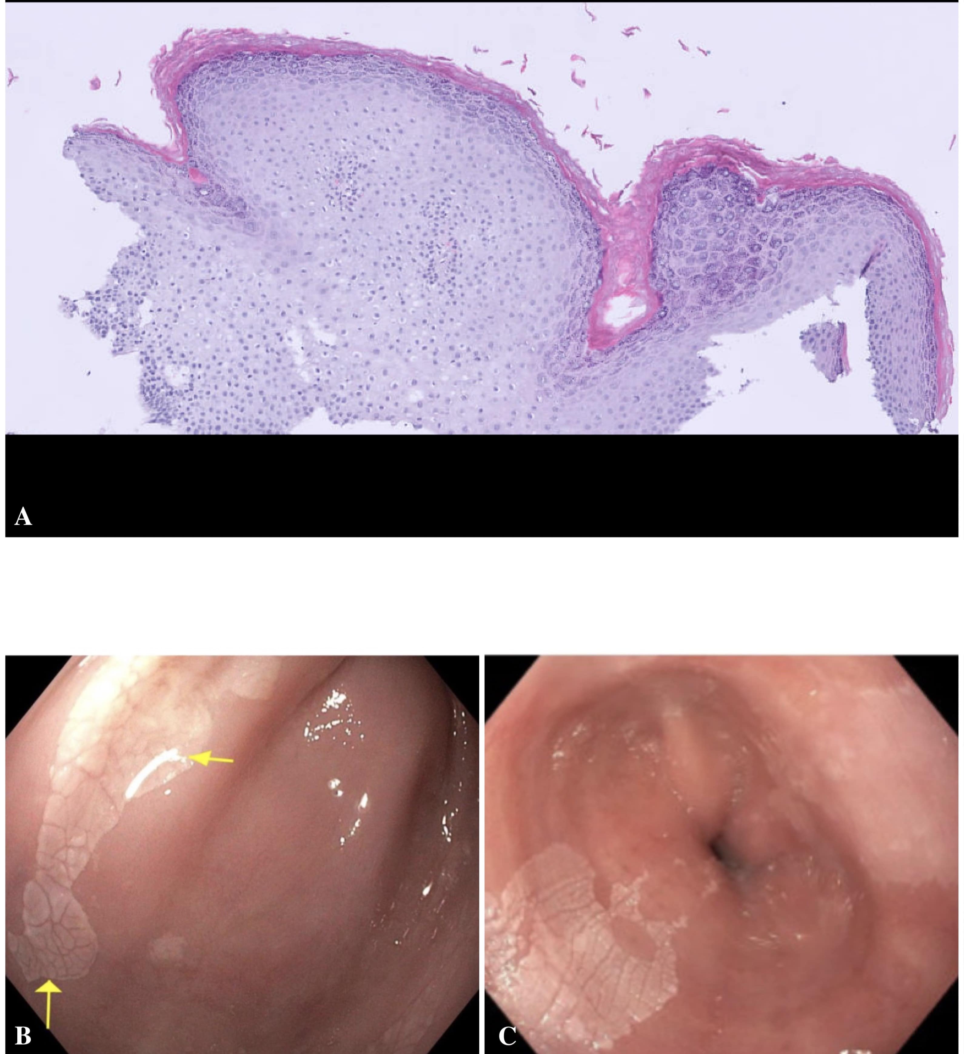
Case Description/Methods: A 62-year-old male with a history of gastroesophageal reflux disease and heavy alcohol use was seen in clinic for dysphagia and food regurgitation. He was started on omeprazole and scheduled for an EGD in two weeks. The EGD revealed multiple white plaques in the middle third and lower third of the esophagus. Biopsies were taken from the esophageal plaques, and pathology revealed epidermoid metaplasia with hyperkeratosis and a prominent granular layer; no dysplasia was identified. A year after the initial EGD, a follow-up EGD showed similar findings. Targeted biopsies were taken from the distal esophageal plaques, and pathology results revealed epidermoid metaplasia. Random biopsies were also taken from the normal-appearing area of the lower third of the esophagus, and pathology results again revealed epidermoid metaplasia. Given the absence of dysplasia or cancer, the decision was made to follow the patient conservatively with a repeat EGD in one year.
Discussion: While these lesions typically exhibit a distinctive gooseflesh appearance and nodular pattern according to the literature, our patient's case revealed a significant finding. Random biopsies taken from the seemingly normal mucosa of the distal esophagus also showed signs of EEM, indicating the presence of microscopic disease that could easily be missed if only targeted biopsies were obtained. Given its rarity, there are no current guidelines on surveillance, the extent of esophageal biopsies that should be obtained, or the best treatment modality. We recommend performing both random and targeted biopsies at regular intervals throughout the mid and distal esophagus to accurately assess the extent of the disease. Additionally, we advise close endoscopic surveillance every 6-12 months, paying particular attention to any subtle changes that could indicate progression to high-grade dysplasia or squamous cell cancer.

Disclosures:
Raakhi Menon, DO, Anirudha Chatterjee, MD, Kanana Aburayyan, MD, Hafiz Ghani, MD, Juliana Yang, MD. P0560 - Unveiling Epidermoid Metaplasia: A Rare Esophageal Transformation, ACG 2024 Annual Scientific Meeting Abstracts. Philadelphia, PA: American College of Gastroenterology.
University of Texas Medical Branch, Galveston, TX
Introduction: Esophageal epidermoid metaplasia (EEM) is a rare premalignant lesion associated with esophageal squamous dysplasia and carcinoma. It is commonly linked to middle-aged women, has a strong association with tobacco and alcohol use, and occurs in less than 2 out of 1,000 consecutive esophageal biopsies. It may present with symptoms like dysphagia and a globus sensation or be asymptomatic and discovered incidentally during esophagogastroduodenoscopy (EGD). Currently, there are no guidelines on surveillance for this high-risk lesion, which makes it difficult to curate a treatment regimen for patients.
Case Description/Methods: A 62-year-old male with a history of gastroesophageal reflux disease and heavy alcohol use was seen in clinic for dysphagia and food regurgitation. He was started on omeprazole and scheduled for an EGD in two weeks. The EGD revealed multiple white plaques in the middle third and lower third of the esophagus. Biopsies were taken from the esophageal plaques, and pathology revealed epidermoid metaplasia with hyperkeratosis and a prominent granular layer; no dysplasia was identified. A year after the initial EGD, a follow-up EGD showed similar findings. Targeted biopsies were taken from the distal esophageal plaques, and pathology results revealed epidermoid metaplasia. Random biopsies were also taken from the normal-appearing area of the lower third of the esophagus, and pathology results again revealed epidermoid metaplasia. Given the absence of dysplasia or cancer, the decision was made to follow the patient conservatively with a repeat EGD in one year.
Discussion: While these lesions typically exhibit a distinctive gooseflesh appearance and nodular pattern according to the literature, our patient's case revealed a significant finding. Random biopsies taken from the seemingly normal mucosa of the distal esophagus also showed signs of EEM, indicating the presence of microscopic disease that could easily be missed if only targeted biopsies were obtained. Given its rarity, there are no current guidelines on surveillance, the extent of esophageal biopsies that should be obtained, or the best treatment modality. We recommend performing both random and targeted biopsies at regular intervals throughout the mid and distal esophagus to accurately assess the extent of the disease. Additionally, we advise close endoscopic surveillance every 6-12 months, paying particular attention to any subtle changes that could indicate progression to high-grade dysplasia or squamous cell cancer.

Figure: Figure 1A. Microscopic Description: Esophageal squamous epithelium with a compact layer of orthokeratosis and a prominent granular layer with keratohyalin granules, resembling the epidermis of the skin.
1B. & C. Endoscopic Description: Localized mucosal changes characterized by whitish discoloration and altered texture in the lower third of the esophagus.
1B. & C. Endoscopic Description: Localized mucosal changes characterized by whitish discoloration and altered texture in the lower third of the esophagus.
Disclosures:
Raakhi Menon indicated no relevant financial relationships.
Anirudha Chatterjee indicated no relevant financial relationships.
Kanana Aburayyan indicated no relevant financial relationships.
Hafiz Ghani indicated no relevant financial relationships.
Juliana Yang indicated no relevant financial relationships.
Raakhi Menon, DO, Anirudha Chatterjee, MD, Kanana Aburayyan, MD, Hafiz Ghani, MD, Juliana Yang, MD. P0560 - Unveiling Epidermoid Metaplasia: A Rare Esophageal Transformation, ACG 2024 Annual Scientific Meeting Abstracts. Philadelphia, PA: American College of Gastroenterology.
