Sunday Poster Session
Category: Endoscopy Video Forum
P0459 - Palliative Use of LAMS in Tandem to Alleviate Malignant Bowel Obstruction in a Patient with Small Bowel Adenocarcinoma with Carcinomatosis
Sunday, October 27, 2024
3:30 PM - 7:00 PM ET
Location: Exhibit Hall E

Has Audio

Ibrahim Yaghnam, MD
Penn State Health Milton S. Hershey Medical Center
Hummelstown, PA
Presenting Author(s)
Smriti Kochhar, DO1, Ibrahim Yaghnam, MD2, Abraham Mathew, MD1
1Penn State Health Milton S. Hershey Medical Center, Hershey, PA; 2Penn State Health Milton S. Hershey Medical Center, Hummelstown, PA
Introduction: The management of patients who present with small bowel obstruction varies depending on the etiology and the comorbidities of the patient. Small bowel adenocarcinoma is a rare tumor of the small bowel, which often presents as small bowel obstruction and is associated with a significant high mortality. While endoscopic management of a focal malignant-related obstruction is well-established, using duodenal stents or endoscopic gastrojejunostomy with lumen-apposing metal stents (LAMS), the endoscopic management of multiple areas of obstruction is not as well documented.
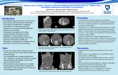
Case Description/Methods: The patient is a 55-year-old woman with a history of small bowel adenocarcinoma with carcinomatosis. She presented with worsening abdominal pain, distention, nausea and vomiting. Imaging demonstrated severe dilation of the small bowel at multiple levels. She was deemed a poor surgical candidate. After careful considerations of the potential life-threatening complications, we decided to pursue placement of tandem lumen-apposing metal stents to palliate the obstructions in the small bowel and colon. We explained to the patient that this is not curative, and it was purely palliative in nature. The patient agreed to undergo the procedure (Video). Using a curvilinear array endoscopic ultrasound, a 20 mm x 10 mm LAMS was placed between the stomach and an area of dilated small bowel. This was sutured in place using an endoscopic suturing system to maintain the gastroenterostomy in place, which was then subsequently dilated to 20 mm using a controlled radial expansion balloon. We then used a combination of direct endoscopy, endoscopic ultrasound, and trans-abdominal ultrasound to identify the next area of LAMS placement for an entero-enterostomy. A 20 mm x 10 mm LAMS was placed between the jejunum and a downstream obstructed portion of small bowel, and it was subsequently dilated to 20 mm. Both areas of small bowel showed diffuse dilation and ischemic changes.
Discussion: Post procedurally, the patient felt dramatic improvement in her symptoms of bloating, nausea and vomiting. Imaging demonstrated decompression of her small bowel. The patient lived an additional three months, until she ultimately expired in her home with her family at her side. She did not require any additional hospitalizations since our procedure.

Disclosures:
Smriti Kochhar, DO1, Ibrahim Yaghnam, MD2, Abraham Mathew, MD1. P0459 - Palliative Use of LAMS in Tandem to Alleviate Malignant Bowel Obstruction in a Patient with Small Bowel Adenocarcinoma with Carcinomatosis, ACG 2024 Annual Scientific Meeting Abstracts. Philadelphia, PA: American College of Gastroenterology.
1Penn State Health Milton S. Hershey Medical Center, Hershey, PA; 2Penn State Health Milton S. Hershey Medical Center, Hummelstown, PA
Introduction: The management of patients who present with small bowel obstruction varies depending on the etiology and the comorbidities of the patient. Small bowel adenocarcinoma is a rare tumor of the small bowel, which often presents as small bowel obstruction and is associated with a significant high mortality. While endoscopic management of a focal malignant-related obstruction is well-established, using duodenal stents or endoscopic gastrojejunostomy with lumen-apposing metal stents (LAMS), the endoscopic management of multiple areas of obstruction is not as well documented.
Case Description/Methods: The patient is a 55-year-old woman with a history of small bowel adenocarcinoma with carcinomatosis. She presented with worsening abdominal pain, distention, nausea and vomiting. Imaging demonstrated severe dilation of the small bowel at multiple levels. She was deemed a poor surgical candidate. After careful considerations of the potential life-threatening complications, we decided to pursue placement of tandem lumen-apposing metal stents to palliate the obstructions in the small bowel and colon. We explained to the patient that this is not curative, and it was purely palliative in nature. The patient agreed to undergo the procedure (Video). Using a curvilinear array endoscopic ultrasound, a 20 mm x 10 mm LAMS was placed between the stomach and an area of dilated small bowel. This was sutured in place using an endoscopic suturing system to maintain the gastroenterostomy in place, which was then subsequently dilated to 20 mm using a controlled radial expansion balloon. We then used a combination of direct endoscopy, endoscopic ultrasound, and trans-abdominal ultrasound to identify the next area of LAMS placement for an entero-enterostomy. A 20 mm x 10 mm LAMS was placed between the jejunum and a downstream obstructed portion of small bowel, and it was subsequently dilated to 20 mm. Both areas of small bowel showed diffuse dilation and ischemic changes.
Discussion: Post procedurally, the patient felt dramatic improvement in her symptoms of bloating, nausea and vomiting. Imaging demonstrated decompression of her small bowel. The patient lived an additional three months, until she ultimately expired in her home with her family at her side. She did not require any additional hospitalizations since our procedure.

Figure: Figure: (Left) Coronal CT image showing dilated loops of small bowel prior to decompression with LAMS. Th coronal CT showing decompressed small bowel after LAMS placement, both the gastro-enteric LAMS (Middle) and entero-enteric LAMS (Right) are seen.
Disclosures:
Smriti Kochhar indicated no relevant financial relationships.
Ibrahim Yaghnam indicated no relevant financial relationships.
Abraham Mathew indicated no relevant financial relationships.
Smriti Kochhar, DO1, Ibrahim Yaghnam, MD2, Abraham Mathew, MD1. P0459 - Palliative Use of LAMS in Tandem to Alleviate Malignant Bowel Obstruction in a Patient with Small Bowel Adenocarcinoma with Carcinomatosis, ACG 2024 Annual Scientific Meeting Abstracts. Philadelphia, PA: American College of Gastroenterology.
