Sunday Poster Session
Category: Biliary/Pancreas
P0075 - Mirizzi Syndrome: A Pain in the Neck (of the Gallbladder)
Sunday, October 27, 2024
3:30 PM - 7:00 PM ET
Location: Exhibit Hall E

Has Audio

Rachael Hagen, DO
University of Connecticut Health
Farmington, CT
Presenting Author(s)
Rachael Hagen, DO1, Minh Thu T.. Nguyen, MD2, Stefan Thorarensen, MD1, Ashwin Pillai, MD1, Neil D.. Parikh, MD3
1University of Connecticut Health, Farmington, CT; 2University of Connecticut Health Center, Farmington, CT; 3Hartford Hospital, Harford, CT
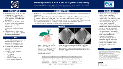
Introduction: Mirizzi syndrome results from the compression of the common hepatic duct (CHD) by gallstones in the cystic duct or Hartmann’s pouch, leading to symptoms of biliary obstruction. Over a span of 23 years, it was identified in 0.18% of all cholecystectomies. Here, we describe a case of Mirizzi syndrome treated with percutaneous cholecystostomy.
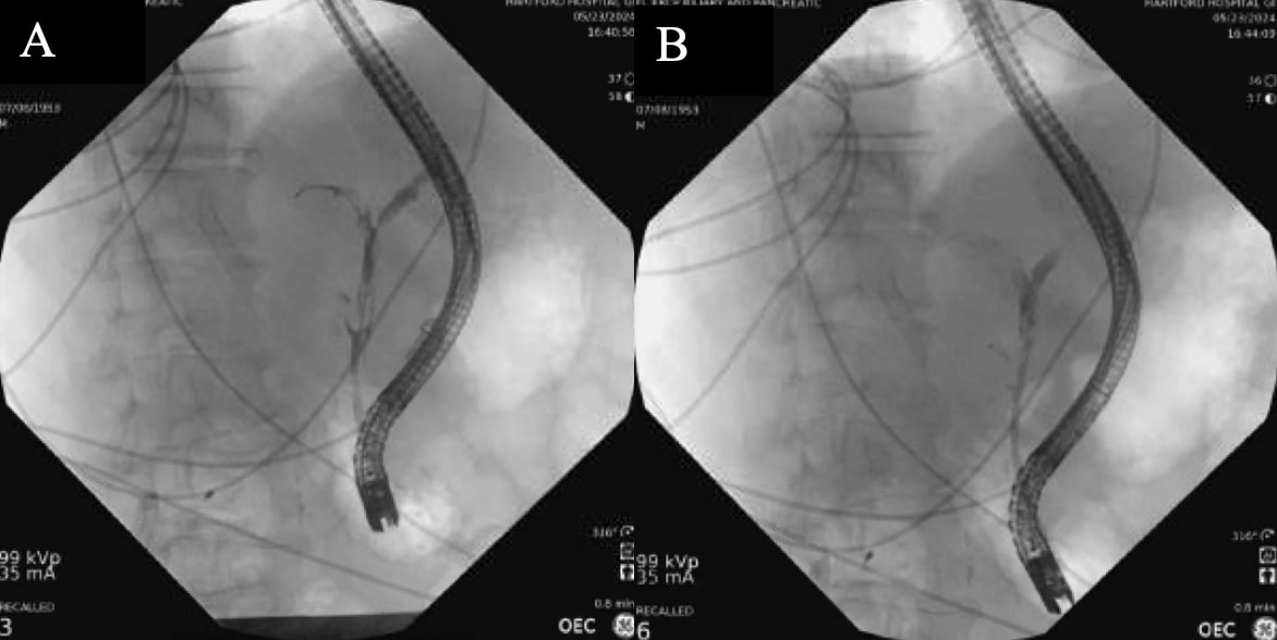
Case Description/Methods: A 70-year-old male was admitted for decompensated heart failure requiring Impella® placement while awaiting heart transplant. He developed epigastric pain associated with nausea and vomiting. Laboratory results showed alkaline phosphatase (440 U/L), aspartate aminotransferase (233 U/L), alanine aminotransferase (289 U/L), and total bilirubin (0.9 mg/dL), with normal lipase. Abdominal computed tomography indicated gallbladder wall thickening with cholelithiasis, consistent with acute cholecystitis. Endoscopic retrograde cholangiopancreatography (ERCP) revealed a cholecystocholedochal fistula in the middle third of the common bile duct (CBD), indicative of Mirizzi syndrome type II, requiring plastic stent placement (Fig. 1). Given his poor candidacy for cholecystectomy, he underwent percutaneous cholecystostomy tube placement and was advised to return for a follow-up ERCP in 3 months for stent removal.
Discussion: Mirizzi syndrome results from gallstones blocking the CHD, potentially causing erosion into the bile duct and the formation of a cholecystocholedochal fistula. Risk factors for Mirizzi syndrome include a tortuous cystic duct, a low insertion of the cystic duct into the CBD, and a thin gallbladder wall. Based off the Csendes classification, the patient was diagnosed with Mirizzi syndrome type II, characterized by the fistula affecting less than one third of the CBD.
Optimal outcomes of Mirizzi syndrome requires timely management and evaluation with ERCP for stone retrieval and stent placement. Early intervention is crucial to mitigate complications, such as bile duct injury. Moreover, inadequate recognition before surgery is associated with high preoperative morbidity and mortality rates. Open cholecystectomy is the gold standard treatment, while laparoscopic surgery is contraindicated. Unfortunately, our patient was unable to undergo surgery due to high-risk comorbidities. There are few reported cases in the literature of patients with Mirizzi syndrome treated with percutaneous cholecystostomy. These cases are limited to elderly patients with severe comorbidities, which largely dictate their prognosis.

Disclosures:
Rachael Hagen, DO1, Minh Thu T.. Nguyen, MD2, Stefan Thorarensen, MD1, Ashwin Pillai, MD1, Neil D.. Parikh, MD3. P0075 - Mirizzi Syndrome: A Pain in the Neck (of the Gallbladder), ACG 2024 Annual Scientific Meeting Abstracts. Philadelphia, PA: American College of Gastroenterology.
1University of Connecticut Health, Farmington, CT; 2University of Connecticut Health Center, Farmington, CT; 3Hartford Hospital, Harford, CT
Introduction: Mirizzi syndrome results from the compression of the common hepatic duct (CHD) by gallstones in the cystic duct or Hartmann’s pouch, leading to symptoms of biliary obstruction. Over a span of 23 years, it was identified in 0.18% of all cholecystectomies. Here, we describe a case of Mirizzi syndrome treated with percutaneous cholecystostomy.
Case Description/Methods: A 70-year-old male was admitted for decompensated heart failure requiring Impella® placement while awaiting heart transplant. He developed epigastric pain associated with nausea and vomiting. Laboratory results showed alkaline phosphatase (440 U/L), aspartate aminotransferase (233 U/L), alanine aminotransferase (289 U/L), and total bilirubin (0.9 mg/dL), with normal lipase. Abdominal computed tomography indicated gallbladder wall thickening with cholelithiasis, consistent with acute cholecystitis. Endoscopic retrograde cholangiopancreatography (ERCP) revealed a cholecystocholedochal fistula in the middle third of the common bile duct (CBD), indicative of Mirizzi syndrome type II, requiring plastic stent placement (Fig. 1). Given his poor candidacy for cholecystectomy, he underwent percutaneous cholecystostomy tube placement and was advised to return for a follow-up ERCP in 3 months for stent removal.
Discussion: Mirizzi syndrome results from gallstones blocking the CHD, potentially causing erosion into the bile duct and the formation of a cholecystocholedochal fistula. Risk factors for Mirizzi syndrome include a tortuous cystic duct, a low insertion of the cystic duct into the CBD, and a thin gallbladder wall. Based off the Csendes classification, the patient was diagnosed with Mirizzi syndrome type II, characterized by the fistula affecting less than one third of the CBD.
Optimal outcomes of Mirizzi syndrome requires timely management and evaluation with ERCP for stone retrieval and stent placement. Early intervention is crucial to mitigate complications, such as bile duct injury. Moreover, inadequate recognition before surgery is associated with high preoperative morbidity and mortality rates. Open cholecystectomy is the gold standard treatment, while laparoscopic surgery is contraindicated. Unfortunately, our patient was unable to undergo surgery due to high-risk comorbidities. There are few reported cases in the literature of patients with Mirizzi syndrome treated with percutaneous cholecystostomy. These cases are limited to elderly patients with severe comorbidities, which largely dictate their prognosis.

Figure: Figure 1a-b. ERCP revealed localized stenosis in the middle third of the CBD measuring 15 mm, consistent with Mirizzi syndrome type II according to the Csendes classification system. One biliary stent with a single external flap was placed, and another with a single internal flap was placed 8 cm into the CBD.
Disclosures:
Rachael Hagen indicated no relevant financial relationships.
Minh Thu Nguyen indicated no relevant financial relationships.
Stefan Thorarensen indicated no relevant financial relationships.
Ashwin Pillai indicated no relevant financial relationships.
Neil Parikh indicated no relevant financial relationships.
Rachael Hagen, DO1, Minh Thu T.. Nguyen, MD2, Stefan Thorarensen, MD1, Ashwin Pillai, MD1, Neil D.. Parikh, MD3. P0075 - Mirizzi Syndrome: A Pain in the Neck (of the Gallbladder), ACG 2024 Annual Scientific Meeting Abstracts. Philadelphia, PA: American College of Gastroenterology.
