Monday Poster Session
Category: Biliary/Pancreas
P1811 - Bouveret Syndrome: A Rare Cause of Gastric Outlet Obstruction
Monday, October 28, 2024
10:30 AM - 4:00 PM ET
Location: Exhibit Hall E

Has Audio

Dimo Dimitrov, MD
St. Elizabeth's Medical Center, Boston University School of Medicine
Boston, MA
Presenting Author(s)
Maram Alenzi, MD1, Dimo Dimitrov, MD1, Iyiad Alabdul Razzak, MD2, Eduardo Vega, MD1, Syed Kashif. Mahmood, MD, MPH2
1St. Elizabeth's Medical Center, Boston University School of Medicine, Boston, MA; 2St. Elizabeth's Medical Center, Boston University School of Medicine, Brighton, MA
Introduction: Bouveret syndrome stands out as a distinct and rare complication of cholelithiasis (1-3%) with high mortality rate ranging from 12-30%. This unique condition arises when a gallstone traverses a cholecystoduodenal fistula, entering the duodenum and subsequently causing gastric outlet obstruction. Delay in diagnosis is associated with a high mortality rate.
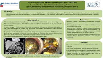
Case Description/Methods: A 66-year-old female with a past medical history of obesity and hyperlipidemia presented with a one-week history of abdominal pain, nausea, and vomiting. On presentation, her vital signs were normal. Her blood work was notable for leukocytosis to 16.2 x 10³ /ul. Liver function test was within normal limit apart from mildly elevated alkaline phosphatase at 114 U/L. Computed tomography (CT) revealed features of acute cholecystitis, cholecystoduodenal fistula, pneumobilia, and multiple gallstones the largest of which was 5-cm in size eroding the duodenal bulb. Also, the stomach was distended on the CT suggestive of gastric outlet obstruction and establishing a diagnosis of Bouveret syndrome. The patient was started on intravenous antibiotics. A total of three endoscopic procedures were performed to fragment the duodenal bulb portion of the stone using electrohydraulic lithotripsy. While lithotripsy successfully cleared the gastric outlet obstruction, the entire stone could not be removed due to its large size. Hence, the patient underwent open gastrotomy with successful removal of several large stones in the duodenal bulb and the cholecystoduodenal fistulous tract. Post-operatively, the patient recovered well and remained asymptomatic. A follow-up esophagogastroduodenoscopy (EGD) six weeks later confirmed the closure of the fistula.
Discussion: Bouveret syndrome is an exceedingly rare complication of gallstone disease diagnosed based on clinical presentation and abdominal imaging findings. Due to its rarity, there is no standardized treatment approach. Typically, endoscopic stone extraction or lithotripsy is preferred due to its safety and minimal invasiveness. However, if endoscopic methods are unsuccessful, surgery may be necessary. Our patient was successfully treated with a combination of antibiotics, therapeutic endoscopy, and exploratory laparotomy. This case highlights the critical role of multidisciplinary team approach to facilitate early diagnosis and the use of both endoscopic and surgical interventions to minimize invasiveness and ensure timely extraction of the obstructing stones.

Disclosures:
Maram Alenzi, MD1, Dimo Dimitrov, MD1, Iyiad Alabdul Razzak, MD2, Eduardo Vega, MD1, Syed Kashif. Mahmood, MD, MPH2. P1811 - Bouveret Syndrome: A Rare Cause of Gastric Outlet Obstruction, ACG 2024 Annual Scientific Meeting Abstracts. Philadelphia, PA: American College of Gastroenterology.
1St. Elizabeth's Medical Center, Boston University School of Medicine, Boston, MA; 2St. Elizabeth's Medical Center, Boston University School of Medicine, Brighton, MA
Introduction: Bouveret syndrome stands out as a distinct and rare complication of cholelithiasis (1-3%) with high mortality rate ranging from 12-30%. This unique condition arises when a gallstone traverses a cholecystoduodenal fistula, entering the duodenum and subsequently causing gastric outlet obstruction. Delay in diagnosis is associated with a high mortality rate.
Case Description/Methods: A 66-year-old female with a past medical history of obesity and hyperlipidemia presented with a one-week history of abdominal pain, nausea, and vomiting. On presentation, her vital signs were normal. Her blood work was notable for leukocytosis to 16.2 x 10³ /ul. Liver function test was within normal limit apart from mildly elevated alkaline phosphatase at 114 U/L. Computed tomography (CT) revealed features of acute cholecystitis, cholecystoduodenal fistula, pneumobilia, and multiple gallstones the largest of which was 5-cm in size eroding the duodenal bulb. Also, the stomach was distended on the CT suggestive of gastric outlet obstruction and establishing a diagnosis of Bouveret syndrome. The patient was started on intravenous antibiotics. A total of three endoscopic procedures were performed to fragment the duodenal bulb portion of the stone using electrohydraulic lithotripsy. While lithotripsy successfully cleared the gastric outlet obstruction, the entire stone could not be removed due to its large size. Hence, the patient underwent open gastrotomy with successful removal of several large stones in the duodenal bulb and the cholecystoduodenal fistulous tract. Post-operatively, the patient recovered well and remained asymptomatic. A follow-up esophagogastroduodenoscopy (EGD) six weeks later confirmed the closure of the fistula.
Discussion: Bouveret syndrome is an exceedingly rare complication of gallstone disease diagnosed based on clinical presentation and abdominal imaging findings. Due to its rarity, there is no standardized treatment approach. Typically, endoscopic stone extraction or lithotripsy is preferred due to its safety and minimal invasiveness. However, if endoscopic methods are unsuccessful, surgery may be necessary. Our patient was successfully treated with a combination of antibiotics, therapeutic endoscopy, and exploratory laparotomy. This case highlights the critical role of multidisciplinary team approach to facilitate early diagnosis and the use of both endoscopic and surgical interventions to minimize invasiveness and ensure timely extraction of the obstructing stones.

Figure: A. CT abdomen/pelvis showing the presence of gallstones and a cholecystoduodenal fistula with a large calcified stone. | B. Endoscopic visualization of the fistulizing stone before lithotripsy. | C. Endoscopic visualization of partial stone fragmentations after lithotripsy.
Disclosures:
Maram Alenzi indicated no relevant financial relationships.
Dimo Dimitrov indicated no relevant financial relationships.
Iyiad Alabdul Razzak indicated no relevant financial relationships.
Eduardo Vega indicated no relevant financial relationships.
Syed Mahmood indicated no relevant financial relationships.
Maram Alenzi, MD1, Dimo Dimitrov, MD1, Iyiad Alabdul Razzak, MD2, Eduardo Vega, MD1, Syed Kashif. Mahmood, MD, MPH2. P1811 - Bouveret Syndrome: A Rare Cause of Gastric Outlet Obstruction, ACG 2024 Annual Scientific Meeting Abstracts. Philadelphia, PA: American College of Gastroenterology.
