Monday Poster Session
Category: Biliary/Pancreas
P1835 - A Rare Case of Pancreatic Head Epidermoid Cyst
Monday, October 28, 2024
10:30 AM - 4:00 PM ET
Location: Exhibit Hall E

Has Audio
- TL
Tooba Laeeq, MD
University of Nevada
Las Vegas, NV
Presenting Author(s)
Tooba Laeeq, MD1, Amith Subhash, MD2, Shahid Wahid, MD2
1University of Nevada, Las Vegas, NV; 2Kirk Kerkorian School of Medicine at the University of Nevada, Las Vegas, NV
Introduction: Epidermoid cysts (EC) in the pancreas are rare. We describe a case of an EC in the head of the pancreas that presented as pancreatitis and small bowel pseudo-obstruction. To our knowledge, this is the first case in the absence of a clear intrapancreatic splenule.
Case Description/Methods: A 49-year-old male with a past medical history of alcohol use disorder presented with generalized abdominal pain, constipation, nausea, and vomiting for one day. Vitals revealed tachycardia and tachypnea. Labs showed BUN 11, hematocrit 54%, alanine aminotransferase 104 IU/L, alkaline phosphatase 113 U/L, lipase 2995 U/L, triglycerides 813 mg/dL, WBC 15.8 k/mm3, . Computed tomography (CT) of the abdomen and pelvis with intravenous contrast showed severe acute pancreatitis, small bowel ileus, and multiple renal cystic lesions. The patient was admitted to the intensive care unit for management of severe pancreatitis and small bowel pseudo-obstruction. The patient’s hospital course was complicated by an infected pseudocyst requiring endoscopic cystogastrostomy and CT-guided percutaneous drainage of additional intraperitoneal fluid collections/abscesses. Cultures from the above fluid collections showed growth of methicillin-sensitive Staphylococcus aureus and Candida albicans. Following treatment with antibacterial and antifungal agents, the patient was ultimately discharged to a skilled nursing facility for ongoing physical therapy. He later returned for outpatient lumen-apposing metal stent removal during which endoscopic ultrasound (US) discovered an atrophic appearing pancreatic head and a 2.2 cm anechoic cyst in the head of the pancreas. A suspected mural nodule measuring 1.3 cm was seen occupying the majority of the cyst cavity. Under US guidance, a targeted fine needle biopsy was performed of the nodule. Pathology revealed a diagnosis of pancreatic EC.
Discussion: Pancreatic EC is characterized by non-neoplastic keratinizing epithelium usually arising from intrapancreatic splenic tissue. The presence of keratinization and the absence of lymphoid components allows them to be differentiated from other kinds of cysts. They are usually discovered in the fourth decade of life. There are three hypotheses regarding the development of EC in the pancreas, namely the mesothelial invagination theory, the lymph space theory, and the endodermal inclusion theory. Diagnosis is often incidental. Due to their potential to mimic malignant lesions, treatment is via surgical resection unless a diagnosis is established.

Disclosures:
Tooba Laeeq, MD1, Amith Subhash, MD2, Shahid Wahid, MD2. P1835 - A Rare Case of Pancreatic Head Epidermoid Cyst, ACG 2024 Annual Scientific Meeting Abstracts. Philadelphia, PA: American College of Gastroenterology.
1University of Nevada, Las Vegas, NV; 2Kirk Kerkorian School of Medicine at the University of Nevada, Las Vegas, NV
Introduction: Epidermoid cysts (EC) in the pancreas are rare. We describe a case of an EC in the head of the pancreas that presented as pancreatitis and small bowel pseudo-obstruction. To our knowledge, this is the first case in the absence of a clear intrapancreatic splenule.
Case Description/Methods: A 49-year-old male with a past medical history of alcohol use disorder presented with generalized abdominal pain, constipation, nausea, and vomiting for one day. Vitals revealed tachycardia and tachypnea. Labs showed BUN 11, hematocrit 54%, alanine aminotransferase 104 IU/L, alkaline phosphatase 113 U/L, lipase 2995 U/L, triglycerides 813 mg/dL, WBC 15.8 k/mm3, . Computed tomography (CT) of the abdomen and pelvis with intravenous contrast showed severe acute pancreatitis, small bowel ileus, and multiple renal cystic lesions. The patient was admitted to the intensive care unit for management of severe pancreatitis and small bowel pseudo-obstruction. The patient’s hospital course was complicated by an infected pseudocyst requiring endoscopic cystogastrostomy and CT-guided percutaneous drainage of additional intraperitoneal fluid collections/abscesses. Cultures from the above fluid collections showed growth of methicillin-sensitive Staphylococcus aureus and Candida albicans. Following treatment with antibacterial and antifungal agents, the patient was ultimately discharged to a skilled nursing facility for ongoing physical therapy. He later returned for outpatient lumen-apposing metal stent removal during which endoscopic ultrasound (US) discovered an atrophic appearing pancreatic head and a 2.2 cm anechoic cyst in the head of the pancreas. A suspected mural nodule measuring 1.3 cm was seen occupying the majority of the cyst cavity. Under US guidance, a targeted fine needle biopsy was performed of the nodule. Pathology revealed a diagnosis of pancreatic EC.
Discussion: Pancreatic EC is characterized by non-neoplastic keratinizing epithelium usually arising from intrapancreatic splenic tissue. The presence of keratinization and the absence of lymphoid components allows them to be differentiated from other kinds of cysts. They are usually discovered in the fourth decade of life. There are three hypotheses regarding the development of EC in the pancreas, namely the mesothelial invagination theory, the lymph space theory, and the endodermal inclusion theory. Diagnosis is often incidental. Due to their potential to mimic malignant lesions, treatment is via surgical resection unless a diagnosis is established.

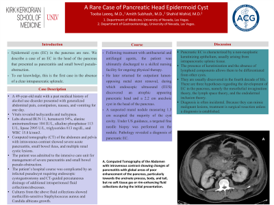
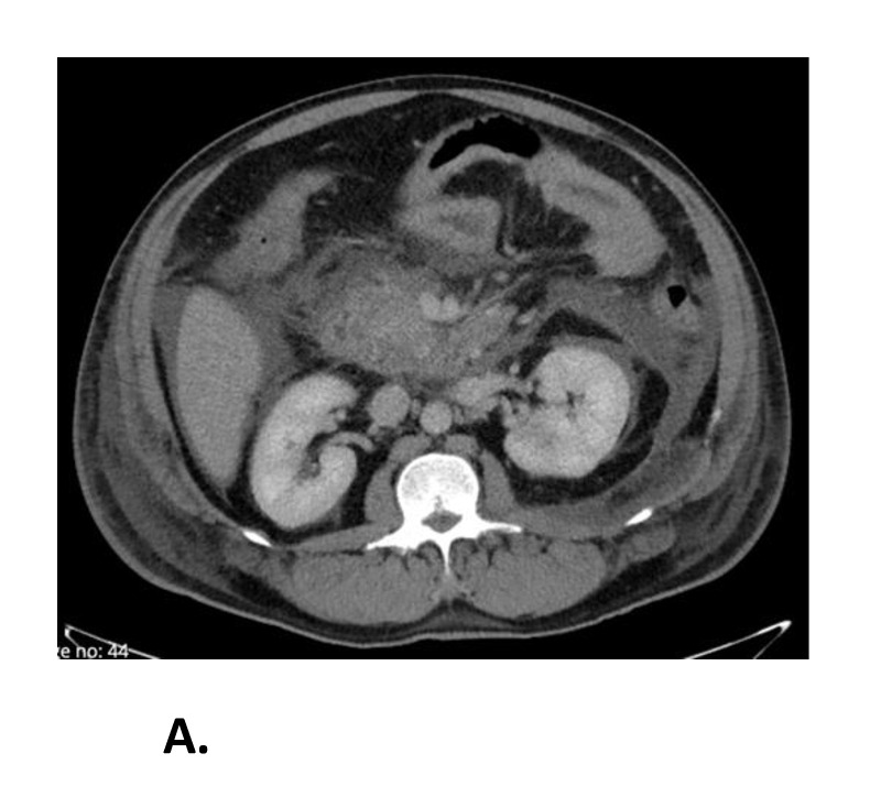
Figure: A. Computed Tomography of the Abdomen with intravenous contrast showing changes of pancreatitis with global areas of poor enhancement of the pancreas, particularly towards the uncinate process, body, and tail but no soft tissue gas or rim-enhancing fluid collections during the initial presentation.
Disclosures:
Tooba Laeeq indicated no relevant financial relationships.
Amith Subhash indicated no relevant financial relationships.
Shahid Wahid indicated no relevant financial relationships.
Tooba Laeeq, MD1, Amith Subhash, MD2, Shahid Wahid, MD2. P1835 - A Rare Case of Pancreatic Head Epidermoid Cyst, ACG 2024 Annual Scientific Meeting Abstracts. Philadelphia, PA: American College of Gastroenterology.

