Monday Poster Session
Category: Biliary/Pancreas
P1838 - A Rare Case of Solitary Fibrous Tumor of the Pancreas
Monday, October 28, 2024
10:30 AM - 4:00 PM ET
Location: Exhibit Hall E

Has Audio

Astin Worden, MD
Mayo Clinic
Phoenix, AZ
Presenting Author(s)
Astin Worden, MD1, Danial Haris. Shaikh, MD2, Michelle Ann. Anderson, MD, MSc3, Zhi Fong, MD1, Rahul Pannala, MD3
1Mayo Clinic, Phoenix, AZ; 2Mayo Clinic, Cave Creek, AZ; 3Mayo Clinic, Scottsdale, AZ
Introduction: Solitary fibrous tumor (SFT) is a rare mesenchymal neoplasm that most often involves the pleura but can occur at various anatomical sites. It is exceedingly rare to find in the pancreas. In the limited cases reported, they present in midlife, equally in men and women, and are typically discovered incidentally or upon workup for vague abdominal symptoms.
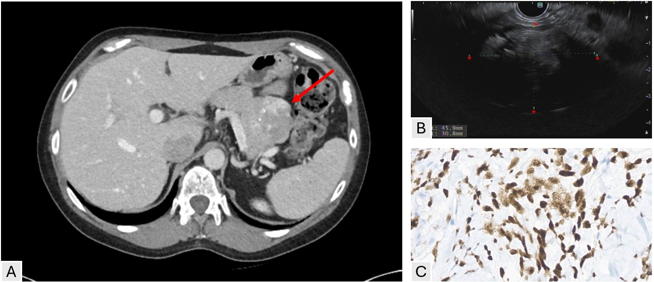
Case Description/Methods: A 56-year-old female with a history of fallopian tube cyst was incidentally found to have a pancreatic tail mass on computed tomography scan (CT) performed for evaluation of lower abdominal pain. The pancreatic lesion was 5 by 6 cm in size with a heterogeneous appearance and scattered calcifications (Image A). She subsequently underwent endoscopic ultrasound, which revealed a hypoechoic heterogenous solid mass in the tail of the pancreas with mild upstream pancreatic ductal dilatation (Image B). Fine-needle biopsy was consistent with SFT staining strongly positive for STAT6 (Image C). A distal pancreatectomy is planned.
Discussion: To date, fewer than 20 cases of pancreatic SFT have been reported in the literature. In these sparse cases, they more commonly occur in the head of the pancreas but can also be found in the tail, as seen in this case. Radiologically, SFTs can mimic more common pancreatic neoplasms such as solid pseudopapillary neoplasms, neuroendocrine tumors, or gastrointestinal stromal tumors. On CT they appear as well-defined, sometimes lobulated masses, isodense to skeletal muscle with heterogeneous contrast enhancement. STAT6 immunohistochemistry is highly sensitive and specific for SFT. The natural history is not well defined given the rarity of this entity, but risk stratification scores centered around tumor size, patient age, mitotic activity, and presence of necrosis have been proposed. Although most pancreatic SFTs have a benign course, some may harbor malignant potential necessitating complete surgical excision as the primary treatment. Long-term follow-up is recommended due to the risk of recurrence or metastasis which can be seen in about 10% of cases.

Disclosures:
Astin Worden, MD1, Danial Haris. Shaikh, MD2, Michelle Ann. Anderson, MD, MSc3, Zhi Fong, MD1, Rahul Pannala, MD3. P1838 - A Rare Case of Solitary Fibrous Tumor of the Pancreas, ACG 2024 Annual Scientific Meeting Abstracts. Philadelphia, PA: American College of Gastroenterology.
1Mayo Clinic, Phoenix, AZ; 2Mayo Clinic, Cave Creek, AZ; 3Mayo Clinic, Scottsdale, AZ
Introduction: Solitary fibrous tumor (SFT) is a rare mesenchymal neoplasm that most often involves the pleura but can occur at various anatomical sites. It is exceedingly rare to find in the pancreas. In the limited cases reported, they present in midlife, equally in men and women, and are typically discovered incidentally or upon workup for vague abdominal symptoms.
Case Description/Methods: A 56-year-old female with a history of fallopian tube cyst was incidentally found to have a pancreatic tail mass on computed tomography scan (CT) performed for evaluation of lower abdominal pain. The pancreatic lesion was 5 by 6 cm in size with a heterogeneous appearance and scattered calcifications (Image A). She subsequently underwent endoscopic ultrasound, which revealed a hypoechoic heterogenous solid mass in the tail of the pancreas with mild upstream pancreatic ductal dilatation (Image B). Fine-needle biopsy was consistent with SFT staining strongly positive for STAT6 (Image C). A distal pancreatectomy is planned.
Discussion: To date, fewer than 20 cases of pancreatic SFT have been reported in the literature. In these sparse cases, they more commonly occur in the head of the pancreas but can also be found in the tail, as seen in this case. Radiologically, SFTs can mimic more common pancreatic neoplasms such as solid pseudopapillary neoplasms, neuroendocrine tumors, or gastrointestinal stromal tumors. On CT they appear as well-defined, sometimes lobulated masses, isodense to skeletal muscle with heterogeneous contrast enhancement. STAT6 immunohistochemistry is highly sensitive and specific for SFT. The natural history is not well defined given the rarity of this entity, but risk stratification scores centered around tumor size, patient age, mitotic activity, and presence of necrosis have been proposed. Although most pancreatic SFTs have a benign course, some may harbor malignant potential necessitating complete surgical excision as the primary treatment. Long-term follow-up is recommended due to the risk of recurrence or metastasis which can be seen in about 10% of cases.

Figure: A) CT showing a 5.0 x 6.0 x 4.0 cm heterogeneous pancreatic tail mass with few scattered internal calcifications. B) EUS with irregular heterogenous mass with focal calcifications in the tail of the pancreas. C) Pathology with tumor cells strongly and diffusely positive for STAT6.
Disclosures:
Astin Worden indicated no relevant financial relationships.
Danial Shaikh indicated no relevant financial relationships.
Michelle Anderson: Boston Scientific – Consultant.
Zhi Fong indicated no relevant financial relationships.
Rahul Pannala: HCL Technologies – Consultant. Nestle Healthsciences – Advisor or Review Panel Member.
Astin Worden, MD1, Danial Haris. Shaikh, MD2, Michelle Ann. Anderson, MD, MSc3, Zhi Fong, MD1, Rahul Pannala, MD3. P1838 - A Rare Case of Solitary Fibrous Tumor of the Pancreas, ACG 2024 Annual Scientific Meeting Abstracts. Philadelphia, PA: American College of Gastroenterology.
