Monday Poster Session
Category: Biliary/Pancreas
P1843 - Pancreatic Neuroendocrine Tumor Masquerades as Adenocarcinoma of Unknown Origin
Monday, October 28, 2024
10:30 AM - 4:00 PM ET
Location: Exhibit Hall E

Has Audio
- JC
Jay C. Clark, DO
Tripler Army Medical Center
Honolulu, HI
Presenting Author(s)
Jay C. Clark, DO, Hema Narlapati, DO, Jeffrey Berenberg, MD, Judy Freeman, MD
Tripler Army Medical Center, Honolulu, HI
Introduction: Pancreatic neuroendocrine tumors are rare and may be classified as either functional or nonfunctional depending on secretion hormones. Most neuroendocrine tumors are indolent and slow growing. A subset of cases are incidentally discovered as metastatic disease rather than a primary mass.
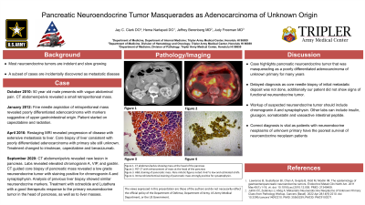
Case Description/Methods: In October 2010, a 50-year-old male presented with vague abdominal pain. CT abdomen/pelvis revealed a small retroperitoneal mass superior to the third portion of duodenum. In January 2012, fine needle aspiration of the mass revealed poorly differentiated adenocarcinoma with cell block showing markers suggestive of upper gastrointestinal origin. The patient was treated with concurrent capecitabine and radiation. In April 2016, restaging MRI revealed disease progression with extensive metastasis to the liver. Core biopsy of the liver was consistent with poorly differentiated adenocarcinoma, with primary site still unknown. He was started on capecitabine and oxaliplatin with a subsequent MRI that showed progression of liver disease. Treatment was switched to irinotecan, capecitabine and bevacizumab with partial response in the liver. In September 2020, CT abdomen/pelvis revealed a new lesion in the pancreas. Labs revealed elevated chromogranin A, VIP, and gastrin. CT guided core biopsy of pancreatic mass revealed a low grade neuroendocrine tumor with staining positive for chromogranin A and synaptophysin. Analysis of previous liver biopsy showed similar neuroendocrine markers. The patient was treated with octreotide and subsequently Lutathera with a good therapeutic response to the primary neuroendocrine tumor in the head of the pancreas, as well as to the liver masses.
Discussion: This case highlights a unique presentation of a pancreatic neuroendocrine tumor that was likely masquerading as a poorly differentiated adenocarcinoma of unknown primary for many years. Proper diagnosis was delayed as core needle biopsy of initial metastatic deposit was not done. Additionally, our patient didn't show clinical signs of a functional neuroendocrine tumor. When suspecting a neuroendocrine tumor, workup should include chromogranin A and synaptophysin. Other lab work such as insulin, glucagon, somatostatin, and vasoactive intestinal peptide can also help identify a functional tumor. Identifying the correct neuroendocrine phenotype is key as patients with neuroendocrine neoplasms of unknown primary have the poorest overall survival of neuroendocrine neoplasm patients.
Disclosures:
Jay C. Clark, DO, Hema Narlapati, DO, Jeffrey Berenberg, MD, Judy Freeman, MD. P1843 - Pancreatic Neuroendocrine Tumor Masquerades as Adenocarcinoma of Unknown Origin, ACG 2024 Annual Scientific Meeting Abstracts. Philadelphia, PA: American College of Gastroenterology.
Tripler Army Medical Center, Honolulu, HI
Introduction: Pancreatic neuroendocrine tumors are rare and may be classified as either functional or nonfunctional depending on secretion hormones. Most neuroendocrine tumors are indolent and slow growing. A subset of cases are incidentally discovered as metastatic disease rather than a primary mass.
Case Description/Methods: In October 2010, a 50-year-old male presented with vague abdominal pain. CT abdomen/pelvis revealed a small retroperitoneal mass superior to the third portion of duodenum. In January 2012, fine needle aspiration of the mass revealed poorly differentiated adenocarcinoma with cell block showing markers suggestive of upper gastrointestinal origin. The patient was treated with concurrent capecitabine and radiation. In April 2016, restaging MRI revealed disease progression with extensive metastasis to the liver. Core biopsy of the liver was consistent with poorly differentiated adenocarcinoma, with primary site still unknown. He was started on capecitabine and oxaliplatin with a subsequent MRI that showed progression of liver disease. Treatment was switched to irinotecan, capecitabine and bevacizumab with partial response in the liver. In September 2020, CT abdomen/pelvis revealed a new lesion in the pancreas. Labs revealed elevated chromogranin A, VIP, and gastrin. CT guided core biopsy of pancreatic mass revealed a low grade neuroendocrine tumor with staining positive for chromogranin A and synaptophysin. Analysis of previous liver biopsy showed similar neuroendocrine markers. The patient was treated with octreotide and subsequently Lutathera with a good therapeutic response to the primary neuroendocrine tumor in the head of the pancreas, as well as to the liver masses.
Discussion: This case highlights a unique presentation of a pancreatic neuroendocrine tumor that was likely masquerading as a poorly differentiated adenocarcinoma of unknown primary for many years. Proper diagnosis was delayed as core needle biopsy of initial metastatic deposit was not done. Additionally, our patient didn't show clinical signs of a functional neuroendocrine tumor. When suspecting a neuroendocrine tumor, workup should include chromogranin A and synaptophysin. Other lab work such as insulin, glucagon, somatostatin, and vasoactive intestinal peptide can also help identify a functional tumor. Identifying the correct neuroendocrine phenotype is key as patients with neuroendocrine neoplasms of unknown primary have the poorest overall survival of neuroendocrine neoplasm patients.
Disclosures:
Jay Clark indicated no relevant financial relationships.
Hema Narlapati indicated no relevant financial relationships.
Jeffrey Berenberg indicated no relevant financial relationships.
Judy Freeman indicated no relevant financial relationships.
Jay C. Clark, DO, Hema Narlapati, DO, Jeffrey Berenberg, MD, Judy Freeman, MD. P1843 - Pancreatic Neuroendocrine Tumor Masquerades as Adenocarcinoma of Unknown Origin, ACG 2024 Annual Scientific Meeting Abstracts. Philadelphia, PA: American College of Gastroenterology.
