Monday Poster Session
Category: Biliary/Pancreas
P1846 - Neuroendocrine Tumor of the Cystic Duct: An Unusual Cause of Acute Cholecystitis
Monday, October 28, 2024
10:30 AM - 4:00 PM ET
Location: Exhibit Hall E

Has Audio
- BC
Bogdan Cristescu, MD
Aurora Healthcare
Green Bay, WI
Presenting Author(s)
Bogdan Cristescu, MD1, Geoffrey Bellini, MD2, Ana Cristescu, 1
1Aurora Healthcare, Green Bay, WI; 2St. Luke's Hospital, Milwaukee, WI
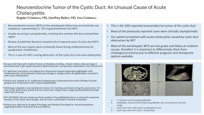
Introduction: Neuroendocrine tumors (NETs) of the extrahepatic biliary tree are extremely rare neoplasms representing 0.2 -2% of gastrointestinal tract NETs, usually occurring in young females and involving the common bile duct and perihilar region. A review of published literature revealed only 9 reported cases of cystic duct NET’s. Most of the case reports were incidentally found during cholecystectomy for symptomatic cholelithiasis. We present a case of a NET causing obstruction of the cystic duct and acute cholecystitis.
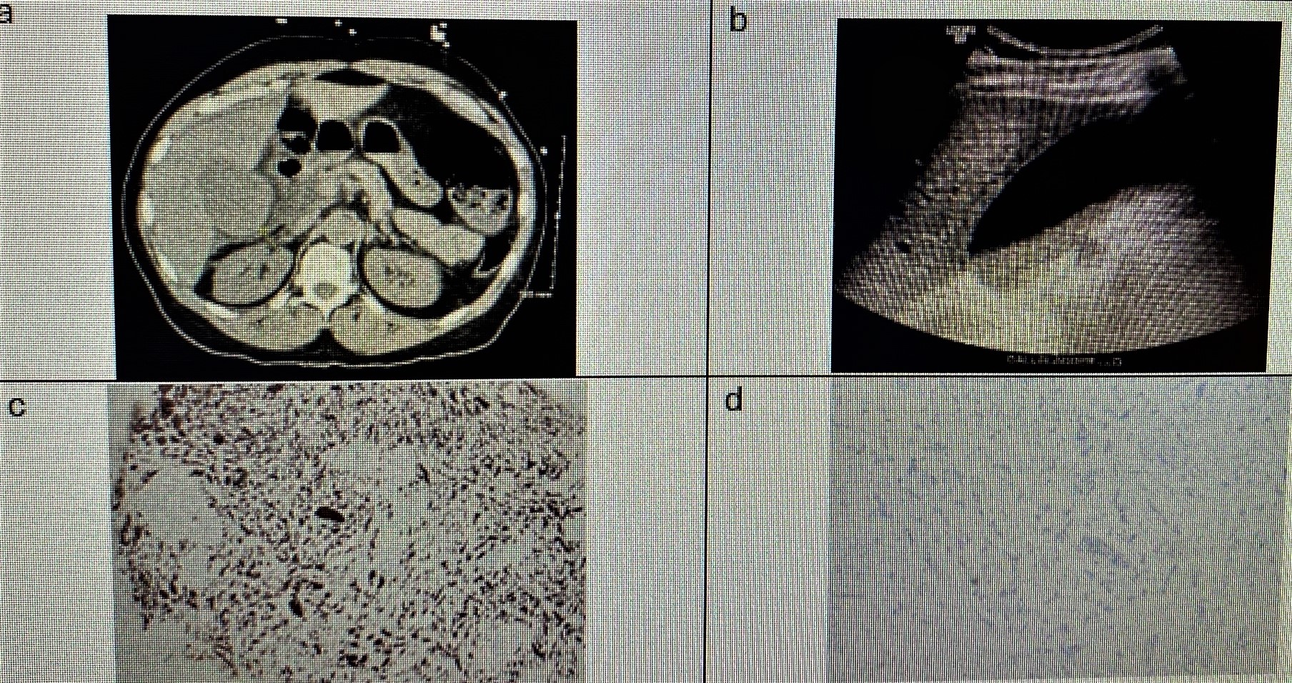
Case Description/Methods: A 63-year-old male with a past medical history of diabetes mellitus, chronic kidney disease stage 3 and GERD presented with right upper quadrant abdominal pain and constipation for 3 days' duration. On physical exam he was tachycardic, tender to palpation in the right upper quadrant of the abdomen. He was found to have leukocytosis and initial LFTs within normal limits. CT abdomen and pelvis showed markedly distended gallbladder with thickened wall and adjacent fat stranding concerning for acute cholecystitis. Abdominal ultrasound confirmed acute cholecystitis with thickened gallbladder wall at 5 mm, pericholecystic fluid and mild to moderate echogenic sludge within the gallbladder. Patient was started on IV antibiotics with piperacillin-tazobactam and underwent laparoscopic cholecystectomy with findings of acute gangrenous cholecystitis with ruptured gallbladder. Pathology revealed a neuroendocrine tumor G1, chromogranin, synaptophysin and CD56 positive with Ki-67 less than 3%, involving and obstructing the cystic duct, 4-5 mm in size with tumor present at the cystic duct margin and a single pericholedochal lymph node free of tumor. PET DOTATATE did not reveal any focal uptake in the gallbladder fossa or along the expected location of the cystic duct margin, and no tracer avid nodal or distant metastasis. The patient was referred to Surgical Oncology and Medical Oncology for recommendations regarding further treatment and surveillance.
Discussion: The present case is the 10th reported NET of the cystic duct. Most of the previously reported cases were clinically asymptomatic, while our patient presented with acute cholecystitis caused by cystic duct obstruction by NET. Most of the extrahepatic NETs are low grade and follow an indolent course, therefore it is important to differentiate them from cholangiocarcinomas due to different prognosis and therapeutic options available.

Disclosures:
Bogdan Cristescu, MD1, Geoffrey Bellini, MD2, Ana Cristescu, 1. P1846 - Neuroendocrine Tumor of the Cystic Duct: An Unusual Cause of Acute Cholecystitis, ACG 2024 Annual Scientific Meeting Abstracts. Philadelphia, PA: American College of Gastroenterology.
1Aurora Healthcare, Green Bay, WI; 2St. Luke's Hospital, Milwaukee, WI
Introduction: Neuroendocrine tumors (NETs) of the extrahepatic biliary tree are extremely rare neoplasms representing 0.2 -2% of gastrointestinal tract NETs, usually occurring in young females and involving the common bile duct and perihilar region. A review of published literature revealed only 9 reported cases of cystic duct NET’s. Most of the case reports were incidentally found during cholecystectomy for symptomatic cholelithiasis. We present a case of a NET causing obstruction of the cystic duct and acute cholecystitis.
Case Description/Methods: A 63-year-old male with a past medical history of diabetes mellitus, chronic kidney disease stage 3 and GERD presented with right upper quadrant abdominal pain and constipation for 3 days' duration. On physical exam he was tachycardic, tender to palpation in the right upper quadrant of the abdomen. He was found to have leukocytosis and initial LFTs within normal limits. CT abdomen and pelvis showed markedly distended gallbladder with thickened wall and adjacent fat stranding concerning for acute cholecystitis. Abdominal ultrasound confirmed acute cholecystitis with thickened gallbladder wall at 5 mm, pericholecystic fluid and mild to moderate echogenic sludge within the gallbladder. Patient was started on IV antibiotics with piperacillin-tazobactam and underwent laparoscopic cholecystectomy with findings of acute gangrenous cholecystitis with ruptured gallbladder. Pathology revealed a neuroendocrine tumor G1, chromogranin, synaptophysin and CD56 positive with Ki-67 less than 3%, involving and obstructing the cystic duct, 4-5 mm in size with tumor present at the cystic duct margin and a single pericholedochal lymph node free of tumor. PET DOTATATE did not reveal any focal uptake in the gallbladder fossa or along the expected location of the cystic duct margin, and no tracer avid nodal or distant metastasis. The patient was referred to Surgical Oncology and Medical Oncology for recommendations regarding further treatment and surveillance.
Discussion: The present case is the 10th reported NET of the cystic duct. Most of the previously reported cases were clinically asymptomatic, while our patient presented with acute cholecystitis caused by cystic duct obstruction by NET. Most of the extrahepatic NETs are low grade and follow an indolent course, therefore it is important to differentiate them from cholangiocarcinomas due to different prognosis and therapeutic options available.

Figure: Figure 1 a. CT abdomen with distended gallbladder, b. Gallbladder ultrasound with thickened gallbladder wall, pericholecystic fluid and sludge, c. Cystic duct tumor with positive synaptophysin stain, d. Cystic duct tumor Ki-67 stain < 3% positivity.
Disclosures:
Bogdan Cristescu indicated no relevant financial relationships.
Geoffrey Bellini indicated no relevant financial relationships.
Ana Cristescu indicated no relevant financial relationships.
Bogdan Cristescu, MD1, Geoffrey Bellini, MD2, Ana Cristescu, 1. P1846 - Neuroendocrine Tumor of the Cystic Duct: An Unusual Cause of Acute Cholecystitis, ACG 2024 Annual Scientific Meeting Abstracts. Philadelphia, PA: American College of Gastroenterology.
