Monday Poster Session
Category: Biliary/Pancreas
P1861 - The Conundrum of MRSA Cholecystitis and Cholestatic Liver Enzyme Elevation
Monday, October 28, 2024
10:30 AM - 4:00 PM ET
Location: Exhibit Hall E

Has Audio

Nathanial Bartosek, MD
Beaumont Health
Royal Oak, MI
Presenting Author(s)
Nathanial Bartosek, MD1, Nazli Begum Ozturk, MD2, Usha Kellampalli, MBBS2, Ketan Rana, MD2
1Beaumont Health, Royal Oak, MI; 2Corewell Health, Royal Oak, MI
Introduction: Methicillin-resistant Staphylococcus aureus (MRSA) is a common cause of healthcare-associated infections, presenting in diverse clinical contexts. Cholecystitis secondary to MRSA is a rare but documented finding. This case highlights the management of a complex patient presenting with MRSA bacteremia and cholecystitis.
Case Description/Methods: A 69-year-old male presented with right upper quadrant pain for five days and sepsis with hypotension, lactic acidosis of 2.2 mmol/L, and elevated liver enzymes in a cholestatic pattern (ALP 799 U/L, AST 73 U/L, ALT 124 U/L). Initial management included fluid resuscitation and piperacillin-tazobactam. Blood cultures returned positive for MRSA, prompting the addition of vancomycin. Ultrasound and CT scan revealed gallbladder distension with pericholecystic fluid concerning for cholecystitis. Cholecystectomy was deferred in favor of percutaneous cholecystectomy, which was completed on the third day of admission; purulent gallbladder aspirate revealed many MRSA on culture. Repeat blood cultures two days following drain placement revealed clearance of MRSA bacteremia.
An Endoscopic Retrograde Cholangiopancreatography (ERCP) was completed on the seventh day of admission due to a continued cholestatic pattern of liver enzymes (ALP 567 U/L, AST 50 U/L, ALT 60 U/L), which revealed no biliary obstruction. A transthoracic echocardiogram did not reveal any vegetation. Given a history of esophageal rupture, a four-week course of daptomycin was preferred over further esophageal instrumentation, and his liver enzymes trended down.
Discussion: In this case, the high concentration of MRSA cultured from the gallbladder, rapid resolution of bacteremia following source control, and lack of other infectious sources suggest primary MRSA cholecystitis. Given the lack of biliary obstruction, the cholestatic pattern likely represented hepatic inflammation and bile stasis mediated through inflammatory cytokines or direct bacterial effects. This case emphasizes the need for thorough diagnostic evaluation, including imaging studies and culture-directed therapy, while highlighting the need for rapid, safe source control.
The rarity of MRSA as a cause of cholecystitis necessitates clinical awareness in cases of suspected cholecystitis with severe systemic manifestations. This patient's cholestatic liver enzyme pattern underscores the diverse hepatic responses to systemic infections and the importance of comprehensive diagnostic evaluation to guide treatment strategies.

Disclosures:
Nathanial Bartosek, MD1, Nazli Begum Ozturk, MD2, Usha Kellampalli, MBBS2, Ketan Rana, MD2. P1861 - The Conundrum of MRSA Cholecystitis and Cholestatic Liver Enzyme Elevation, ACG 2024 Annual Scientific Meeting Abstracts. Philadelphia, PA: American College of Gastroenterology.
1Beaumont Health, Royal Oak, MI; 2Corewell Health, Royal Oak, MI
Introduction: Methicillin-resistant Staphylococcus aureus (MRSA) is a common cause of healthcare-associated infections, presenting in diverse clinical contexts. Cholecystitis secondary to MRSA is a rare but documented finding. This case highlights the management of a complex patient presenting with MRSA bacteremia and cholecystitis.
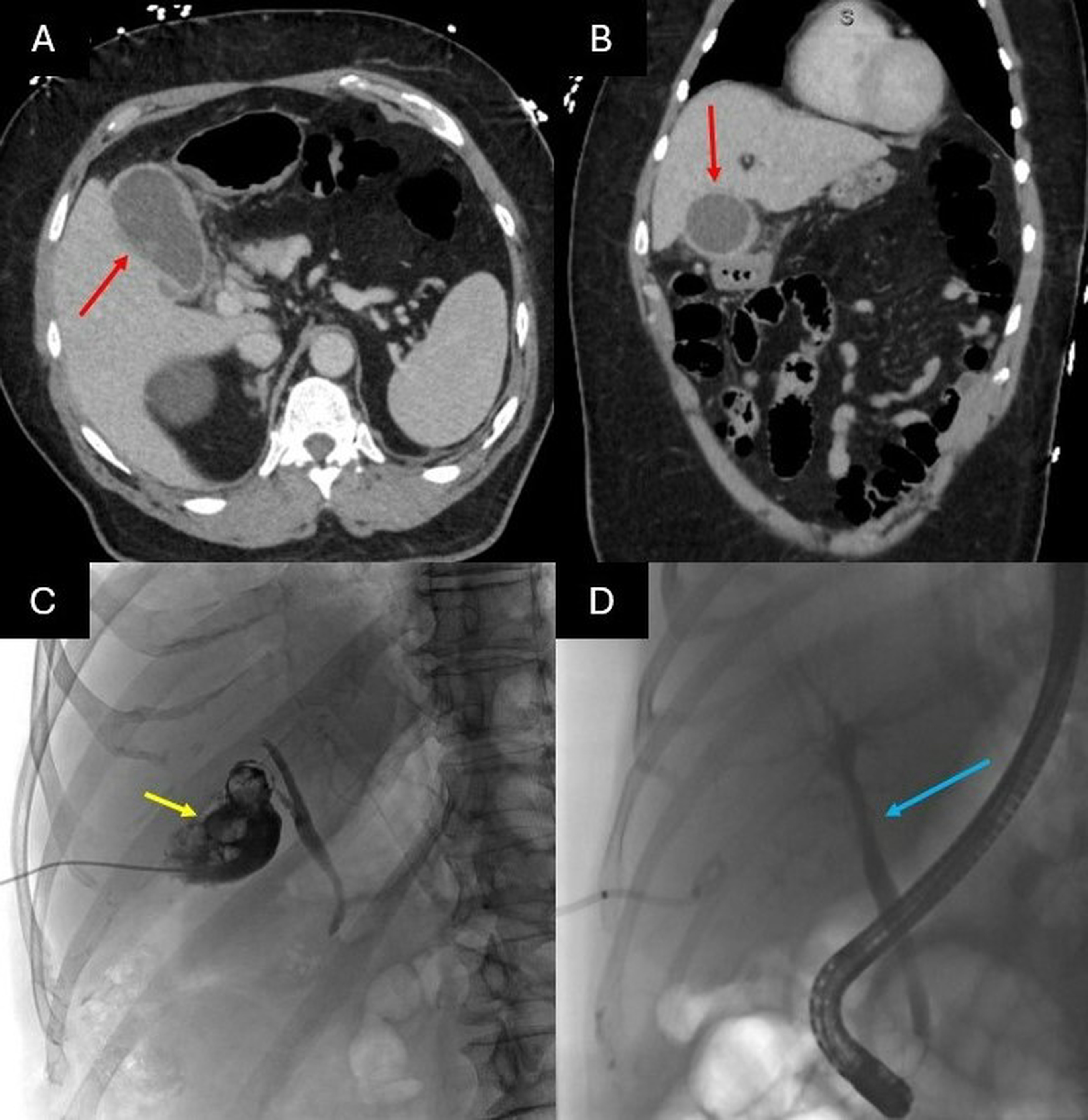
Case Description/Methods: A 69-year-old male presented with right upper quadrant pain for five days and sepsis with hypotension, lactic acidosis of 2.2 mmol/L, and elevated liver enzymes in a cholestatic pattern (ALP 799 U/L, AST 73 U/L, ALT 124 U/L). Initial management included fluid resuscitation and piperacillin-tazobactam. Blood cultures returned positive for MRSA, prompting the addition of vancomycin. Ultrasound and CT scan revealed gallbladder distension with pericholecystic fluid concerning for cholecystitis. Cholecystectomy was deferred in favor of percutaneous cholecystectomy, which was completed on the third day of admission; purulent gallbladder aspirate revealed many MRSA on culture. Repeat blood cultures two days following drain placement revealed clearance of MRSA bacteremia.
An Endoscopic Retrograde Cholangiopancreatography (ERCP) was completed on the seventh day of admission due to a continued cholestatic pattern of liver enzymes (ALP 567 U/L, AST 50 U/L, ALT 60 U/L), which revealed no biliary obstruction. A transthoracic echocardiogram did not reveal any vegetation. Given a history of esophageal rupture, a four-week course of daptomycin was preferred over further esophageal instrumentation, and his liver enzymes trended down.
Discussion: In this case, the high concentration of MRSA cultured from the gallbladder, rapid resolution of bacteremia following source control, and lack of other infectious sources suggest primary MRSA cholecystitis. Given the lack of biliary obstruction, the cholestatic pattern likely represented hepatic inflammation and bile stasis mediated through inflammatory cytokines or direct bacterial effects. This case emphasizes the need for thorough diagnostic evaluation, including imaging studies and culture-directed therapy, while highlighting the need for rapid, safe source control.
The rarity of MRSA as a cause of cholecystitis necessitates clinical awareness in cases of suspected cholecystitis with severe systemic manifestations. This patient's cholestatic liver enzyme pattern underscores the diverse hepatic responses to systemic infections and the importance of comprehensive diagnostic evaluation to guide treatment strategies.

Figure: Figure: (A,B) Computed Tomography (CT) images of noted significant gallbladder distension (Red Arrows). (C) Fluoroscopic images obtained during percutaneous cholecystostomy tube placement, in which multiple filling defects (Yellow Arrow) were appreciated and contrast was shown to progress through the cystic and common bile ducts. (D) Fluoroscopic images during ERCP, displaying a normal cholangiogram with no occlusion appreciated (Blue Arrow).
Disclosures:
Nathanial Bartosek indicated no relevant financial relationships.
Nazli Begum Ozturk indicated no relevant financial relationships.
Usha Kellampalli indicated no relevant financial relationships.
Ketan Rana indicated no relevant financial relationships.
Nathanial Bartosek, MD1, Nazli Begum Ozturk, MD2, Usha Kellampalli, MBBS2, Ketan Rana, MD2. P1861 - The Conundrum of MRSA Cholecystitis and Cholestatic Liver Enzyme Elevation, ACG 2024 Annual Scientific Meeting Abstracts. Philadelphia, PA: American College of Gastroenterology.

