Monday Poster Session
Category: Biliary/Pancreas
P1883 - It Is Not Cholecystitis, or Is It? Look Again: A Rare Incidence of Emphysematous Cholecystitis With Negative Initial Imagining
Monday, October 28, 2024
10:30 AM - 4:00 PM ET
Location: Exhibit Hall E

Has Audio
- JS
Judy Sheffeh, MD
Ascension Macomb-Oakland Hospital
Warren, MI
Presenting Author(s)
Judy Sheffeh, MD, Mitchell Oliver, DO, Katie Claxton, MS, Rita Rehana, MD, Mohammed Barawi, MD
Ascension Macomb-Oakland Hospital, Warren, MI
Introduction: Differential diagnosis for upper abdominal pain is vast. Thorough history-taking, physical exam and tailored diagnostic work up are crucial for accurate diagnosis and management. This is a rare occurrence of emphysematous cholecystitis (EC) that was undetected initially despite the use of gold-standard imaging techniques.
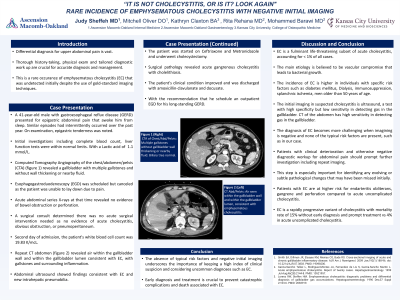
Case Description/Methods: A 41-year-old male presented for epigastric abdominal pain. Computed tomography angiography of the abdomen revealed a gallbladder with multiple gallstones without wall thickening, and no bowel obstruction or perforation. Lactic acid of 1.1. A surgical consult determined there was no acute surgical intervention needed. The pain was believed to be related to stool burden noted on imaging. The patient received laxatives and continued to be treated symptomatically. On the second day of admission, a repeat CT abdomen was performed due to leukocytosis of 19K and intractable severe abdominal pain. The scan revealed air within the gallbladder wall and lumen consistent with EC, accompanied by gallstones and surrounding inflammation. Abdominal ultrasound showed findings consistent with EC and new intrahepatic pneumobilia. The patient was started on antibiotics and underwent cholecystectomy. Surgical pathology revealed acute gangrenous cholecystitis with cholelithiasis. The patient’s clinical condition improved and was discharged with amoxicillin-clavulanate and docusate.
Discussion: EC is a fulminant life-threatening subset of acute cholecystitis, accounting for < 1% of all cases. The incidence of EC is higher in individuals with specific risk factors such as diabetes mellitus, dialysis, immunosuppression, splanchnic ischemia, and in men older than 50 years of age. The initial imaging in suspected cholecystitis is ultrasound, a test with high specificity but low sensitivity in detecting gas in the gallbladder. CT has high sensitivity in detecting gas in the gallbladder. The diagnosis of EC becomes more challenging when imagining is negative and none of the typical risk factors are present, such as in our case. Patients with clinical deterioration and otherwise negative diagnostic workup for abdominal pain should prompt further investigation including repeat imaging. This step is especially important for identifying any evolving or subtle pathological changes that may have been missed initially. EC is a rapidly progressive variant of cholecystitis with mortality rate of 15% without early diagnosis and prompt treatment vs 4% in acute uncomplicated cholecystitis.
Disclosures:
Judy Sheffeh, MD, Mitchell Oliver, DO, Katie Claxton, MS, Rita Rehana, MD, Mohammed Barawi, MD. P1883 - It Is Not Cholecystitis, or Is It? Look Again: A Rare Incidence of Emphysematous Cholecystitis With Negative Initial Imagining, ACG 2024 Annual Scientific Meeting Abstracts. Philadelphia, PA: American College of Gastroenterology.
Ascension Macomb-Oakland Hospital, Warren, MI
Introduction: Differential diagnosis for upper abdominal pain is vast. Thorough history-taking, physical exam and tailored diagnostic work up are crucial for accurate diagnosis and management. This is a rare occurrence of emphysematous cholecystitis (EC) that was undetected initially despite the use of gold-standard imaging techniques.
Case Description/Methods: A 41-year-old male presented for epigastric abdominal pain. Computed tomography angiography of the abdomen revealed a gallbladder with multiple gallstones without wall thickening, and no bowel obstruction or perforation. Lactic acid of 1.1. A surgical consult determined there was no acute surgical intervention needed. The pain was believed to be related to stool burden noted on imaging. The patient received laxatives and continued to be treated symptomatically. On the second day of admission, a repeat CT abdomen was performed due to leukocytosis of 19K and intractable severe abdominal pain. The scan revealed air within the gallbladder wall and lumen consistent with EC, accompanied by gallstones and surrounding inflammation. Abdominal ultrasound showed findings consistent with EC and new intrahepatic pneumobilia. The patient was started on antibiotics and underwent cholecystectomy. Surgical pathology revealed acute gangrenous cholecystitis with cholelithiasis. The patient’s clinical condition improved and was discharged with amoxicillin-clavulanate and docusate.
Discussion: EC is a fulminant life-threatening subset of acute cholecystitis, accounting for < 1% of all cases. The incidence of EC is higher in individuals with specific risk factors such as diabetes mellitus, dialysis, immunosuppression, splanchnic ischemia, and in men older than 50 years of age. The initial imaging in suspected cholecystitis is ultrasound, a test with high specificity but low sensitivity in detecting gas in the gallbladder. CT has high sensitivity in detecting gas in the gallbladder. The diagnosis of EC becomes more challenging when imagining is negative and none of the typical risk factors are present, such as in our case. Patients with clinical deterioration and otherwise negative diagnostic workup for abdominal pain should prompt further investigation including repeat imaging. This step is especially important for identifying any evolving or subtle pathological changes that may have been missed initially. EC is a rapidly progressive variant of cholecystitis with mortality rate of 15% without early diagnosis and prompt treatment vs 4% in acute uncomplicated cholecystitis.
Disclosures:
Judy Sheffeh indicated no relevant financial relationships.
Mitchell Oliver indicated no relevant financial relationships.
Katie Claxton indicated no relevant financial relationships.
Rita Rehana indicated no relevant financial relationships.
Mohammed Barawi indicated no relevant financial relationships.
Judy Sheffeh, MD, Mitchell Oliver, DO, Katie Claxton, MS, Rita Rehana, MD, Mohammed Barawi, MD. P1883 - It Is Not Cholecystitis, or Is It? Look Again: A Rare Incidence of Emphysematous Cholecystitis With Negative Initial Imagining, ACG 2024 Annual Scientific Meeting Abstracts. Philadelphia, PA: American College of Gastroenterology.
