Monday Poster Session
Category: Biliary/Pancreas
P1887 - Case Report: Rare Case of Ewing’s Sarcoma in the Pancreas
Monday, October 28, 2024
10:30 AM - 4:00 PM ET
Location: Exhibit Hall E

Has Audio

Nitin Pendyala, MD
South Brooklyn Health
Brooklyn, NY
Presenting Author(s)
Joseph Choi, DO, Nitin Pendyala, MD, Nikisha Pandya, MD, Syed Karim, MD, Christopher Chum, DO
South Brooklyn Health, Brooklyn, NY
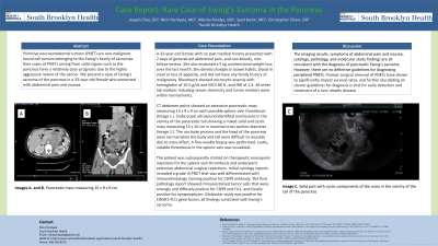
Introduction: Primitive neuroectodermal tumors (PNET) are rare malignant round cell tumors belonging to the Ewing’s family of sarcomas. Rare cases of PNETs arising from solid organs such as the pancreas have a relatively poor prognosis due to the highly aggressive nature of the cancer. We present a case of Ewing’s sarcoma of the pancreas in a 33-year-old female who presented with abdominal pain and nausea.
Case Description/Methods: A 33-year-old female with no past medical history presented with 2 days of generalized abdominal pain, and non-bloody, non-bilious emesis. She also endorsed a 5 kg unintentional weight loss over the last month. She denied changes in bowel habits, blood in stool or loss of appetite, and did not have any family history of malignancy. Bloodwork showed microcytic anemia with hemoglobin of 10.5 g/dL and MCV 80 fL, and INR of 1.3. All other lab markers including serum chemistry and tumor markers were within normal limits.
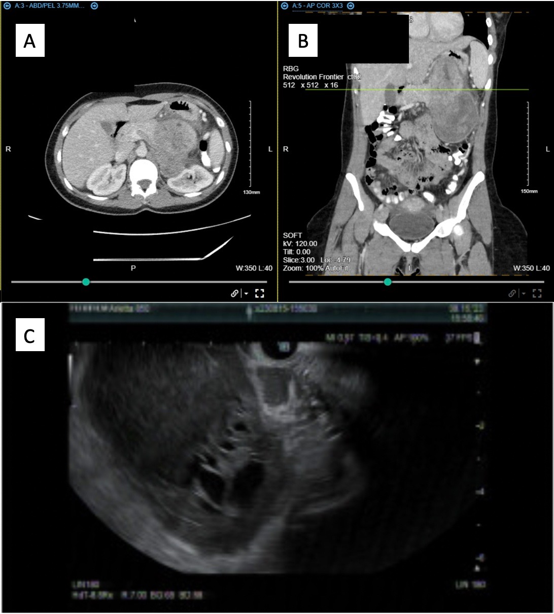
CT abdomen pelvis showed an extensive pancreatic mass measuring 15 x 9 x 9 cm with possible splenic vein thrombosis (Images A. and B.). Endoscopic ultrasound identified oval lesions in the vicinity of the pancreatic tail showing a mixed solid and cystic mass measuring 15 x 10 cm in maximal cross-section diameter (Image C.). The uncinate process and the head of the pancreas were normal while the body and tail were difficult to visualize due to mass effect. A fine-needle biopsy was performed. Lastly, notable thrombosis in the splenic vein was visualized.
The patient subsequently began therapeutic enoxaparin injections for the splenic vein thrombosis and underwent extensive abdominal surgical resections. Initial cytology reports revealed a grade III PNET that was well differentiated with immunohistology staining positive for CD99 antibody. The final pathology report showed immunostained tumor cells that were strongly and diffusely positive for CD99 and FLI1, and focally positive for synaptophysin. Molecular study was positive for EWSR1-FLI1 gene fusion, all findings consistent with Ewing’s sarcoma.
Discussion: The imaging results, symptoms of abdominal pain and nausea, cytology, pathology, and molecular study findings are all consistent with the diagnosis of pancreatic Ewing’s sarcoma. However, there are no definitive guidelines for diagnosing peripheral PNETs. Prompt surgical removal of PNETs have shown to significantly impact survival rates, and further elucidating on clearer guidelines for diagnosis is vital for early detection and treatment of a rare, deadly disease.

Disclosures:
Joseph Choi, DO, Nitin Pendyala, MD, Nikisha Pandya, MD, Syed Karim, MD, Christopher Chum, DO. P1887 - Case Report: Rare Case of Ewing’s Sarcoma in the Pancreas, ACG 2024 Annual Scientific Meeting Abstracts. Philadelphia, PA: American College of Gastroenterology.
South Brooklyn Health, Brooklyn, NY
Introduction: Primitive neuroectodermal tumors (PNET) are rare malignant round cell tumors belonging to the Ewing’s family of sarcomas. Rare cases of PNETs arising from solid organs such as the pancreas have a relatively poor prognosis due to the highly aggressive nature of the cancer. We present a case of Ewing’s sarcoma of the pancreas in a 33-year-old female who presented with abdominal pain and nausea.
Case Description/Methods: A 33-year-old female with no past medical history presented with 2 days of generalized abdominal pain, and non-bloody, non-bilious emesis. She also endorsed a 5 kg unintentional weight loss over the last month. She denied changes in bowel habits, blood in stool or loss of appetite, and did not have any family history of malignancy. Bloodwork showed microcytic anemia with hemoglobin of 10.5 g/dL and MCV 80 fL, and INR of 1.3. All other lab markers including serum chemistry and tumor markers were within normal limits.
CT abdomen pelvis showed an extensive pancreatic mass measuring 15 x 9 x 9 cm with possible splenic vein thrombosis (Images A. and B.). Endoscopic ultrasound identified oval lesions in the vicinity of the pancreatic tail showing a mixed solid and cystic mass measuring 15 x 10 cm in maximal cross-section diameter (Image C.). The uncinate process and the head of the pancreas were normal while the body and tail were difficult to visualize due to mass effect. A fine-needle biopsy was performed. Lastly, notable thrombosis in the splenic vein was visualized.
The patient subsequently began therapeutic enoxaparin injections for the splenic vein thrombosis and underwent extensive abdominal surgical resections. Initial cytology reports revealed a grade III PNET that was well differentiated with immunohistology staining positive for CD99 antibody. The final pathology report showed immunostained tumor cells that were strongly and diffusely positive for CD99 and FLI1, and focally positive for synaptophysin. Molecular study was positive for EWSR1-FLI1 gene fusion, all findings consistent with Ewing’s sarcoma.
Discussion: The imaging results, symptoms of abdominal pain and nausea, cytology, pathology, and molecular study findings are all consistent with the diagnosis of pancreatic Ewing’s sarcoma. However, there are no definitive guidelines for diagnosing peripheral PNETs. Prompt surgical removal of PNETs have shown to significantly impact survival rates, and further elucidating on clearer guidelines for diagnosis is vital for early detection and treatment of a rare, deadly disease.

Figure: Images A. and B. Pancreatic mass measuring 15 x 9 x 9 cm
Image C. Solid part with cystic components of the mass in the vicinity of the tail of the pancreas
Image C. Solid part with cystic components of the mass in the vicinity of the tail of the pancreas
Disclosures:
Joseph Choi indicated no relevant financial relationships.
Nitin Pendyala indicated no relevant financial relationships.
Nikisha Pandya indicated no relevant financial relationships.
Syed Karim indicated no relevant financial relationships.
Christopher Chum indicated no relevant financial relationships.
Joseph Choi, DO, Nitin Pendyala, MD, Nikisha Pandya, MD, Syed Karim, MD, Christopher Chum, DO. P1887 - Case Report: Rare Case of Ewing’s Sarcoma in the Pancreas, ACG 2024 Annual Scientific Meeting Abstracts. Philadelphia, PA: American College of Gastroenterology.
