Monday Poster Session
Category: Biliary/Pancreas
P1888 - Pancreatic Neuroendocrine Tumor causing Duodenal Varices: A Case Report
Monday, October 28, 2024
10:30 AM - 4:00 PM ET
Location: Exhibit Hall E

Has Audio

Tahne Vongsavath, DO
Kirk Kirkorian School of Medicine
Las Vegas, NV
Presenting Author(s)
Tahne Vongsavath, DO, Vignan Manne, MD
Kirk Kirkorian School of Medicine, Las Vegas, NV
Introduction: While gastric and esophageal varices are well described as sequela of portal hypertension, duodenal varices are a rarer presentation. Generally present at the bulb or second part of the duodenum, they have massive bleed and rebleed potential and because of the scarcity of data on them, optimum treatment is not well described. Emerging techniques, including balloon-occluded retrograde obliteration and social assisted obliteration techniques have been described.
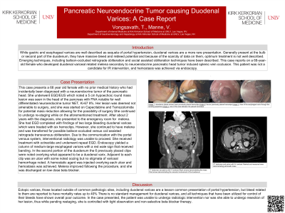
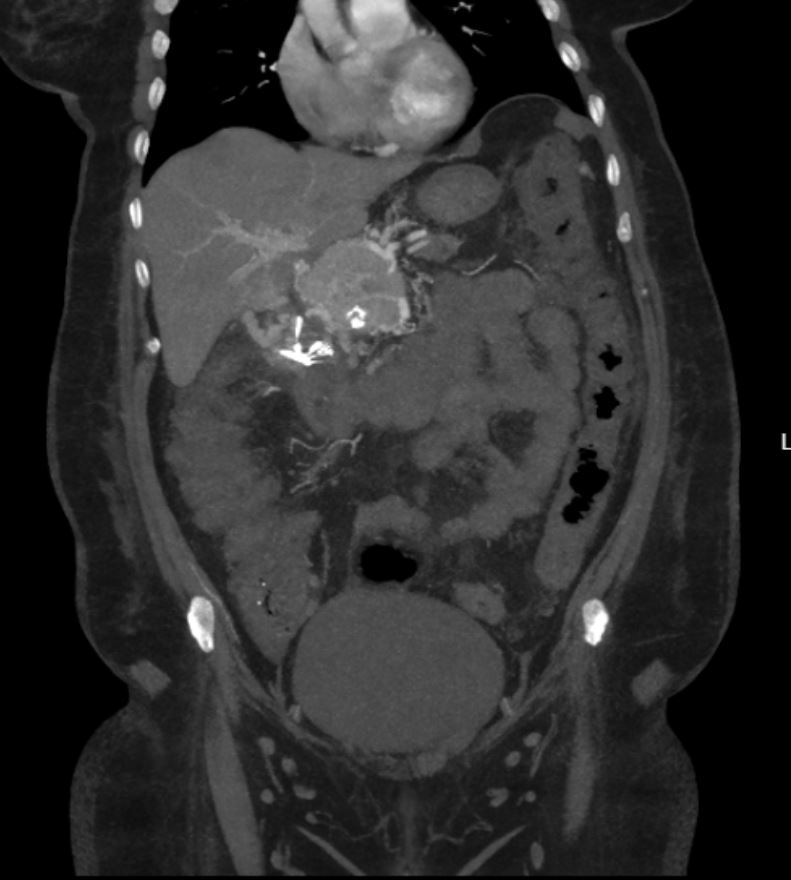
Case Description/Methods: This case presents a 68-year-old female with no prior medical history who was found to have a pancreatic head mass on CT imaging during work up for elevated liver enzymes. She underwent esophogastroduodenoscopy with endoscopic ultrasound that noted a5 cm hypoechoic lesion in the head of the pancreas causing pancreatic duct obstruction. A plastic biliary stent was placed traversing the mass and samples were taken. Fine needle aspiration results reported a well differentiated neuroendocrine tumor Ki-67 9% Her lesion was deemed not amenable to surgery, she was started on Capecitabine and Temozolomide, and was continually restaged in hopes for future intervention. About 2 years following diagnosis she began having melena. She underwent an EGD showing two large bleeding duodenal varices that did not resolve with clipping. She was transferred to a facility with balloon vs coil assisted retrograde transvenous obliteration capability, however due to the communication with the portal venous system, radiology elected not to proceed. She had repeat EGD that noted an esophageal varix and oozing at the duodenal varices in the second part of the duodenum. Hemostasis was achieved with hemostatic peptide gel injection. Melena improved following the procedure, and she was discharged on low dose carvedilol. The patient remains on carvedilol to reduce portal pressure while she continues treatment and restaging.
Discussion: Ectopic varices, like duodenal varices are a lesson common presentation of portal hypertension, but bleeds are reported to have mortality rates up to 40% There is no standard management for duodenal varices, and all techniques that have been utilized for management have shown overall poor outcome. In this case, the patient was unable to undergo radiologic intervention nor was she able to undergo resection of her lesion, thus while pending restaging, she is controlled with observation and carvedilol therapy.

Disclosures:
Tahne Vongsavath, DO, Vignan Manne, MD. P1888 - Pancreatic Neuroendocrine Tumor causing Duodenal Varices: A Case Report, ACG 2024 Annual Scientific Meeting Abstracts. Philadelphia, PA: American College of Gastroenterology.
Kirk Kirkorian School of Medicine, Las Vegas, NV
Introduction: While gastric and esophageal varices are well described as sequela of portal hypertension, duodenal varices are a rarer presentation. Generally present at the bulb or second part of the duodenum, they have massive bleed and rebleed potential and because of the scarcity of data on them, optimum treatment is not well described. Emerging techniques, including balloon-occluded retrograde obliteration and social assisted obliteration techniques have been described.
Case Description/Methods: This case presents a 68-year-old female with no prior medical history who was found to have a pancreatic head mass on CT imaging during work up for elevated liver enzymes. She underwent esophogastroduodenoscopy with endoscopic ultrasound that noted a5 cm hypoechoic lesion in the head of the pancreas causing pancreatic duct obstruction. A plastic biliary stent was placed traversing the mass and samples were taken. Fine needle aspiration results reported a well differentiated neuroendocrine tumor Ki-67 9% Her lesion was deemed not amenable to surgery, she was started on Capecitabine and Temozolomide, and was continually restaged in hopes for future intervention. About 2 years following diagnosis she began having melena. She underwent an EGD showing two large bleeding duodenal varices that did not resolve with clipping. She was transferred to a facility with balloon vs coil assisted retrograde transvenous obliteration capability, however due to the communication with the portal venous system, radiology elected not to proceed. She had repeat EGD that noted an esophageal varix and oozing at the duodenal varices in the second part of the duodenum. Hemostasis was achieved with hemostatic peptide gel injection. Melena improved following the procedure, and she was discharged on low dose carvedilol. The patient remains on carvedilol to reduce portal pressure while she continues treatment and restaging.
Discussion: Ectopic varices, like duodenal varices are a lesson common presentation of portal hypertension, but bleeds are reported to have mortality rates up to 40% There is no standard management for duodenal varices, and all techniques that have been utilized for management have shown overall poor outcome. In this case, the patient was unable to undergo radiologic intervention nor was she able to undergo resection of her lesion, thus while pending restaging, she is controlled with observation and carvedilol therapy.

Figure: Pancreatic head mass
Disclosures:
Tahne Vongsavath indicated no relevant financial relationships.
Vignan Manne indicated no relevant financial relationships.
Tahne Vongsavath, DO, Vignan Manne, MD. P1888 - Pancreatic Neuroendocrine Tumor causing Duodenal Varices: A Case Report, ACG 2024 Annual Scientific Meeting Abstracts. Philadelphia, PA: American College of Gastroenterology.
