Monday Poster Session
Category: Biliary/Pancreas
P1908 - Management of a Complicated Post-Cholecystectomy Course as a Result of Mirizzi Syndrome
Monday, October 28, 2024
10:30 AM - 4:00 PM ET
Location: Exhibit Hall E

Has Audio

Monica A. Huynh, DO
HCA Florida Largo Hospital
Largo, FL
Presenting Author(s)
Monica A. Huynh Williams, DO, Ashley Long, DO, Kevin Yeroushalmi, MD, Meir Mizrahi, MD
HCA Florida Largo Hospital, Largo, FL
Introduction: Mirizzi Syndrome is an obstruction of the common hepatic duct (CHD) due to compression from a stone in the cystic duct or gallbladder.1 Standard treatment is surgical intervention; however, we discuss a patient with Mirizzi syndrome who faced numerous complications postoperatively.
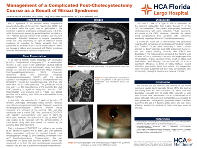
Case Description/Methods: A 55-year-old female presented with obstructive jaundice. Imaging revealed a 2 cm gallstone in the cystic duct causing CHD compression and proximal biliary duct dilation (Image 1,2) consistent with Mirizzi syndrome. Endoscopy with fine needle aspiration and stent placement showed negative pathology and only provided temporary symptomatic relief. Subsequent robotic cholecystectomy showed the gallbladder had eroded into over two-thirds of the common bile duct (CBD) circumference resulting in significant biliary duct distortion. CBD reconstruction and T-tube placement proximal to the existing stent were performed, and both were removed in the following months.
The patient presented for jaundice a year later, and imaging showed proximal CHD occlusion. Initial plans for endoscopic stenting and papillary sphincterotomy were hindered by the inability to traverse a proximal CBD obstruction (Image 3). A transhepatic biliary drain was placed but removed prematurely by the patient. The patient ultimately underwent Roux-en-Y hepaticojejunostomy, complicated by biliary leak suspected to be from the percutaneous transhepatic insertion site, but was managed conservatively.
Discussion: The patient underwent appropriate treatment for a type III Mirizzi syndrome but faced complications requiring a Roux-en-Y hepaticojejunostomy.1 T-tube insertion and removal have the potential to cause biliary duct damage, as witnessed with our patient.2 Comparison of biliary stent and T-tube outcomes concluded that stenting was superior to T-tube placement, offering decreased complications, operative times, lengths of stay, and readmission rates.2 This highlights an opportunity for management guidelines to transition away from the use of T-tubes to utilize safer endoscopic methods of biliary drainage, such as stenting.
References

Disclosures:
Monica A. Huynh Williams, DO, Ashley Long, DO, Kevin Yeroushalmi, MD, Meir Mizrahi, MD. P1908 - Management of a Complicated Post-Cholecystectomy Course as a Result of Mirizzi Syndrome, ACG 2024 Annual Scientific Meeting Abstracts. Philadelphia, PA: American College of Gastroenterology.
HCA Florida Largo Hospital, Largo, FL
Introduction: Mirizzi Syndrome is an obstruction of the common hepatic duct (CHD) due to compression from a stone in the cystic duct or gallbladder.1 Standard treatment is surgical intervention; however, we discuss a patient with Mirizzi syndrome who faced numerous complications postoperatively.
Case Description/Methods: A 55-year-old female presented with obstructive jaundice. Imaging revealed a 2 cm gallstone in the cystic duct causing CHD compression and proximal biliary duct dilation (Image 1,2) consistent with Mirizzi syndrome. Endoscopy with fine needle aspiration and stent placement showed negative pathology and only provided temporary symptomatic relief. Subsequent robotic cholecystectomy showed the gallbladder had eroded into over two-thirds of the common bile duct (CBD) circumference resulting in significant biliary duct distortion. CBD reconstruction and T-tube placement proximal to the existing stent were performed, and both were removed in the following months.
The patient presented for jaundice a year later, and imaging showed proximal CHD occlusion. Initial plans for endoscopic stenting and papillary sphincterotomy were hindered by the inability to traverse a proximal CBD obstruction (Image 3). A transhepatic biliary drain was placed but removed prematurely by the patient. The patient ultimately underwent Roux-en-Y hepaticojejunostomy, complicated by biliary leak suspected to be from the percutaneous transhepatic insertion site, but was managed conservatively.
Discussion: The patient underwent appropriate treatment for a type III Mirizzi syndrome but faced complications requiring a Roux-en-Y hepaticojejunostomy.1 T-tube insertion and removal have the potential to cause biliary duct damage, as witnessed with our patient.2 Comparison of biliary stent and T-tube outcomes concluded that stenting was superior to T-tube placement, offering decreased complications, operative times, lengths of stay, and readmission rates.2 This highlights an opportunity for management guidelines to transition away from the use of T-tubes to utilize safer endoscopic methods of biliary drainage, such as stenting.
References
Valderrama-Treviño AI, Granados-Romero JJ, Espejel-Deloiza M, et al. Updates in Mirizzi syndrome. Hepatobiliary Surg Nutr. 2017;6(3):170-178. doi:10.21037/hbsn.2016.11.01
Young M, Mehta D. Percutaneous Transhepatic Cholangiogram. [Updated 2023 Jul 25]. In: StatPearls [Internet]. Treasure Island (FL): StatPearls Publishing; 2024 Jan-. Available from: https://www.ncbi.nlm.nih.gov/books/NBK493190/

Figure: Image 1 (Left): Computerized tomography of the abdomen/pelvis axial view of large 2 cm gallstone in the cystic duct.
Image 2 (Middle): Computerized tomography of the abdomen/pelvis coronal view of large 2 cm gallstone in the cystic duct with extrahepatic and intrahepatic biliary duct dilation.
Image 3 (Right): Cholangioscopy images of the guidewire coiling as a result of proximal CBD obstruction.
Image 2 (Middle): Computerized tomography of the abdomen/pelvis coronal view of large 2 cm gallstone in the cystic duct with extrahepatic and intrahepatic biliary duct dilation.
Image 3 (Right): Cholangioscopy images of the guidewire coiling as a result of proximal CBD obstruction.
Disclosures:
Monica Huynh Williams indicated no relevant financial relationships.
Ashley Long indicated no relevant financial relationships.
Kevin Yeroushalmi indicated no relevant financial relationships.
Meir Mizrahi indicated no relevant financial relationships.
Monica A. Huynh Williams, DO, Ashley Long, DO, Kevin Yeroushalmi, MD, Meir Mizrahi, MD. P1908 - Management of a Complicated Post-Cholecystectomy Course as a Result of Mirizzi Syndrome, ACG 2024 Annual Scientific Meeting Abstracts. Philadelphia, PA: American College of Gastroenterology.
