Monday Poster Session
Category: Colon
P1964 - A Rare Case of Secondary Perianal Paget's Disease
Monday, October 28, 2024
10:30 AM - 4:00 PM ET
Location: Exhibit Hall E

Has Audio

Colin Westman, DO, MS
Hackensack Meridian Health - Palisades Medical Center
Hackensack, NJ
Presenting Author(s)
Colin Westman, DO, MS1, Vignesh Krishnan Nagesh, MBBS2, Shawn Philip, DO3, Davood K Hosseini, MD3, Nazir Ahmed, MD3, Kunchang Song, MD4, John Sotiriadis, MD, PhD, FACG2
1Hackensack Meridian Health - Palisades Medical Center, Hackensack, NJ; 2Hackensack Meridian Palisades Medical Center, North Bergen, NJ; 3Hackensack University Medical Center, Hackensack, NJ; 4Hackensack Meridian Health - Palisades Medical Center, North Bergen, NJ
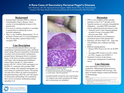
Introduction: Perianal Paget’s Disease (PPD) is a form of Extramammary Paget’s Disease consisting of intraepithelial invasion of the perianal skin. It can present as a primary neoplasm of the perianal skin or as secondary spread from an underlying anorectal invasive carcinoma. The condition is exceedingly rare, representing less than 1% of anal disease and 1.3% of all Paget’s Disease. Herein, we provide a case report of a patient with secondary PPD.
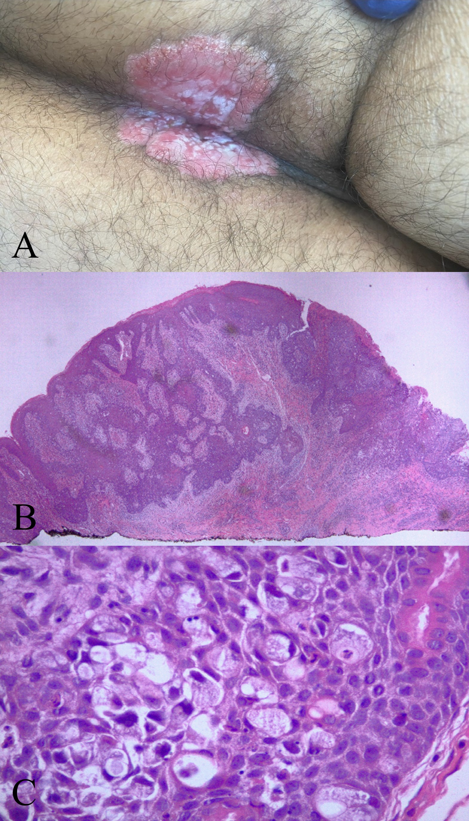
Case Description/Methods: A 50 year old male presented three years ago complaining of perianal pruritus, pain, and blood when wiping. Physical examination of the patient revealed a well-circumscribed, erythematous perianal lesion. Subsequent elective incisional biopsy of the lesion revealed a polypoid lesion with tumor cells containing intracytoplasmic mucin (Figure 1B and 1C). These stained positive for CK7, CK20, CDX2 and negative for GCDFP-15, consistent with secondary PPD. Further workup with and computed tomography was unrevealing for synchronous lesions. The patient was then lost to follow-up until he presented to the gastroenterology office this year with reports of anal pruritus and bleeding. On examination, there was an excoriated, erythematous plaque spreading 4 cm circumferentially around the anal canal (Figure 1A). Repeat exam under anesthesia for biopsy and a colonoscopy are scheduled.
Discussion: PPD is a rare condition, most often diagnosed in the 6th and 7th decades of life and usually presents as a primary intraepithelial malignancy. Secondary PPD, which is associated with an underlying anorectal malignancy, is much rarer. In a prior review of the National Cancer Center Hospital database, 14 cases of secondary PPD were identified between 2006 – 2021. It is typically described as an erythematous, eczematous lesion involving the perianal skin. Histology shows characteristic Paget cells, which are large cells with abundant, pale cytoplasm and large nuclei. In primary PPD, cells are positive for CK7 and GCDFP-15. In secondary PPD cells are positive for CK7, CK20, and CDX2, but negative for GCDFP-15. The management of PPD is poorly defined due to the lack of data. It usually includes excision of the underlying skin lesion as well as evaluation for the spread of malignancy, both endoscopically and radiographically. This case raises awareness for inclusion of PPD in the differential diagnosis under the appropriate circumstances.

Disclosures:
Colin Westman, DO, MS1, Vignesh Krishnan Nagesh, MBBS2, Shawn Philip, DO3, Davood K Hosseini, MD3, Nazir Ahmed, MD3, Kunchang Song, MD4, John Sotiriadis, MD, PhD, FACG2. P1964 - A Rare Case of Secondary Perianal Paget's Disease, ACG 2024 Annual Scientific Meeting Abstracts. Philadelphia, PA: American College of Gastroenterology.
1Hackensack Meridian Health - Palisades Medical Center, Hackensack, NJ; 2Hackensack Meridian Palisades Medical Center, North Bergen, NJ; 3Hackensack University Medical Center, Hackensack, NJ; 4Hackensack Meridian Health - Palisades Medical Center, North Bergen, NJ
Introduction: Perianal Paget’s Disease (PPD) is a form of Extramammary Paget’s Disease consisting of intraepithelial invasion of the perianal skin. It can present as a primary neoplasm of the perianal skin or as secondary spread from an underlying anorectal invasive carcinoma. The condition is exceedingly rare, representing less than 1% of anal disease and 1.3% of all Paget’s Disease. Herein, we provide a case report of a patient with secondary PPD.
Case Description/Methods: A 50 year old male presented three years ago complaining of perianal pruritus, pain, and blood when wiping. Physical examination of the patient revealed a well-circumscribed, erythematous perianal lesion. Subsequent elective incisional biopsy of the lesion revealed a polypoid lesion with tumor cells containing intracytoplasmic mucin (Figure 1B and 1C). These stained positive for CK7, CK20, CDX2 and negative for GCDFP-15, consistent with secondary PPD. Further workup with and computed tomography was unrevealing for synchronous lesions. The patient was then lost to follow-up until he presented to the gastroenterology office this year with reports of anal pruritus and bleeding. On examination, there was an excoriated, erythematous plaque spreading 4 cm circumferentially around the anal canal (Figure 1A). Repeat exam under anesthesia for biopsy and a colonoscopy are scheduled.
Discussion: PPD is a rare condition, most often diagnosed in the 6th and 7th decades of life and usually presents as a primary intraepithelial malignancy. Secondary PPD, which is associated with an underlying anorectal malignancy, is much rarer. In a prior review of the National Cancer Center Hospital database, 14 cases of secondary PPD were identified between 2006 – 2021. It is typically described as an erythematous, eczematous lesion involving the perianal skin. Histology shows characteristic Paget cells, which are large cells with abundant, pale cytoplasm and large nuclei. In primary PPD, cells are positive for CK7 and GCDFP-15. In secondary PPD cells are positive for CK7, CK20, and CDX2, but negative for GCDFP-15. The management of PPD is poorly defined due to the lack of data. It usually includes excision of the underlying skin lesion as well as evaluation for the spread of malignancy, both endoscopically and radiographically. This case raises awareness for inclusion of PPD in the differential diagnosis under the appropriate circumstances.

Figure: Figure 1A. Gross appearance of lesion. 1B. Polypoid squamous lesion, H&E x2 magnification. 1C. Carcinoma cells with abundant foamy cytoplasm, either singly or in small clusters , H&E x40 magnification.
Disclosures:
Colin Westman indicated no relevant financial relationships.
Vignesh Krishnan Nagesh indicated no relevant financial relationships.
Shawn Philip indicated no relevant financial relationships.
Davood K Hosseini indicated no relevant financial relationships.
Nazir Ahmed indicated no relevant financial relationships.
Kunchang Song indicated no relevant financial relationships.
John Sotiriadis: AbbVie – Speakers Bureau.
Colin Westman, DO, MS1, Vignesh Krishnan Nagesh, MBBS2, Shawn Philip, DO3, Davood K Hosseini, MD3, Nazir Ahmed, MD3, Kunchang Song, MD4, John Sotiriadis, MD, PhD, FACG2. P1964 - A Rare Case of Secondary Perianal Paget's Disease, ACG 2024 Annual Scientific Meeting Abstracts. Philadelphia, PA: American College of Gastroenterology.
