Monday Poster Session
Category: Colon
P1966 - Colorectal Lymphangioma: Case Report and Diagnostic Challenges
Monday, October 28, 2024
10:30 AM - 4:00 PM ET
Location: Exhibit Hall E

Has Audio

Dimo Dimitrov, MD
St. Elizabeth's Medical Center, Boston University School of Medicine
Boston, MA
Presenting Author(s)
Dimo Dimitrov, MD1, Jad Mitri, MD1, Sandeep Krishnan, MD, PhD2
1St. Elizabeth's Medical Center, Boston University School of Medicine, Boston, MA; 2St. Elizabeth's Medical Center, Boston University School of Medicine, Brighton, MA
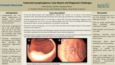
Introduction: Lymphangiomas are rare, benign, hamartomatous malformations of lymphatic tissue, typically found in children, mainly in the head and neck. Colorectal lymphangiomas are very rare and often linked to inflammation, degeneration, or trauma when found in adults. The diagnosis is challenging due to its varied presentation, from incidental imaging findings to acute abdomen, and its rarity. While non-malignant, symptomatic cases may require resection, with a generally good prognosis.
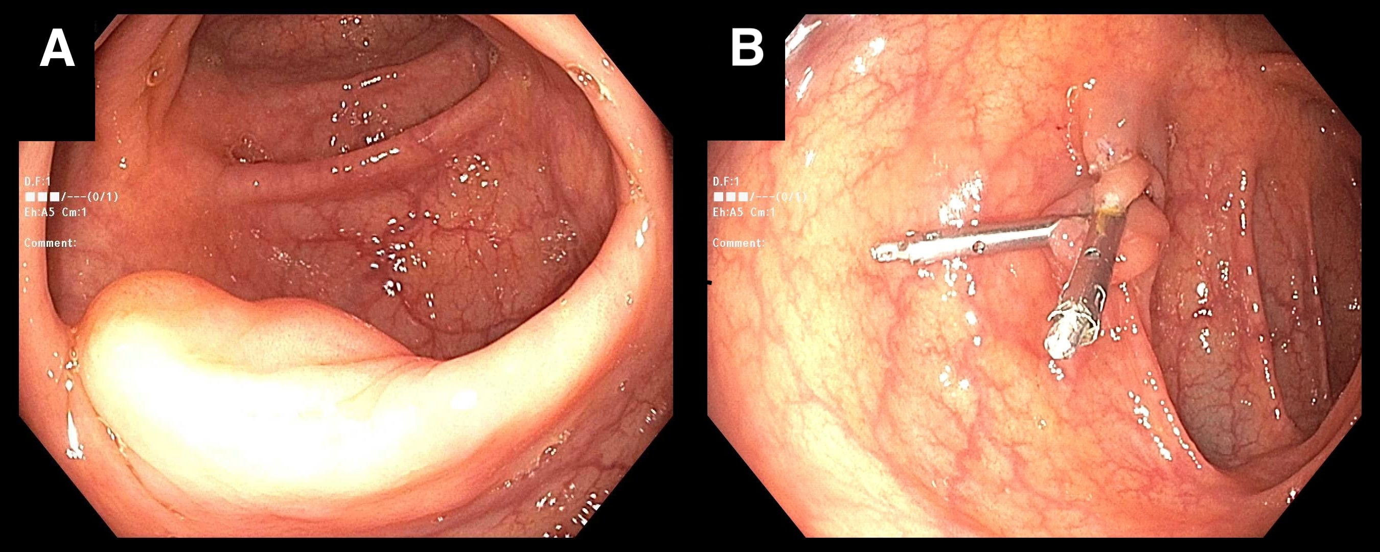
Case Description/Methods: A 57-year-old female patient with hyperlipidemia, obesity, and a known subepithelial colonic mass presented for a surveillance colonoscopy. Seven years ago, a colonoscopy revealed a 1.5 cm subepithelial lesion in the proximal transverse colon. A CT scan then showed a 2.6 cm intraluminal cystic lesion, which reduced to 1.3 cm a year later. The differential diagnoses included benign lesions such as lymphangioma, duplication cyst, and mucocele. As the patient was asymptomatic, no further intervention was planned, and she was scheduled for a surveillance colonoscopy. During the current colonoscopy, a 1.2 cm sessile lesion was found at the hepatic flexure in the proximal transverse colon. Endoscopic mucosal resection with lifting agent injection and snare technique was performed, followed by hemostatic clip placement and India ink tattooing for future reference. Pathology analysis identified a subepithelial lymphangioma.
Discussion: Colorectal lymphangioma, primarily affects men in their 40s to 60s, with 1.4-2:1 male to female distribution. It is most commonly found in the cecum, transverse, and ascending colon. It can vary in size and histology, with the cystic subtype being the most common. Clinically, lymphangiomas are often asymptomatic and discovered incidentally during endoscopy or radiologic studies. They are frequently misdiagnosed until tissue samples are obtained. Initial evaluation typically involves ultrasound, CT, or MRI. Endoscopic ultrasound can provide precise information about the layer of origin and contour of the subepithelial tumor. Colonoscopy provides opportunity for direct visualization and tissue sampling. Close surveillance or endoscopic therapy may be appropriate for asymptomatic lesions smaller than 2.5 cm in diameter. Surgical intervention is considered for larger lesions or in patients who develop complications such as intestinal obstruction, bleeding, intussusception, rupture, or malignant transformation.

Disclosures:
Dimo Dimitrov, MD1, Jad Mitri, MD1, Sandeep Krishnan, MD, PhD2. P1966 - Colorectal Lymphangioma: Case Report and Diagnostic Challenges, ACG 2024 Annual Scientific Meeting Abstracts. Philadelphia, PA: American College of Gastroenterology.
1St. Elizabeth's Medical Center, Boston University School of Medicine, Boston, MA; 2St. Elizabeth's Medical Center, Boston University School of Medicine, Brighton, MA
Introduction: Lymphangiomas are rare, benign, hamartomatous malformations of lymphatic tissue, typically found in children, mainly in the head and neck. Colorectal lymphangiomas are very rare and often linked to inflammation, degeneration, or trauma when found in adults. The diagnosis is challenging due to its varied presentation, from incidental imaging findings to acute abdomen, and its rarity. While non-malignant, symptomatic cases may require resection, with a generally good prognosis.
Case Description/Methods: A 57-year-old female patient with hyperlipidemia, obesity, and a known subepithelial colonic mass presented for a surveillance colonoscopy. Seven years ago, a colonoscopy revealed a 1.5 cm subepithelial lesion in the proximal transverse colon. A CT scan then showed a 2.6 cm intraluminal cystic lesion, which reduced to 1.3 cm a year later. The differential diagnoses included benign lesions such as lymphangioma, duplication cyst, and mucocele. As the patient was asymptomatic, no further intervention was planned, and she was scheduled for a surveillance colonoscopy. During the current colonoscopy, a 1.2 cm sessile lesion was found at the hepatic flexure in the proximal transverse colon. Endoscopic mucosal resection with lifting agent injection and snare technique was performed, followed by hemostatic clip placement and India ink tattooing for future reference. Pathology analysis identified a subepithelial lymphangioma.
Discussion: Colorectal lymphangioma, primarily affects men in their 40s to 60s, with 1.4-2:1 male to female distribution. It is most commonly found in the cecum, transverse, and ascending colon. It can vary in size and histology, with the cystic subtype being the most common. Clinically, lymphangiomas are often asymptomatic and discovered incidentally during endoscopy or radiologic studies. They are frequently misdiagnosed until tissue samples are obtained. Initial evaluation typically involves ultrasound, CT, or MRI. Endoscopic ultrasound can provide precise information about the layer of origin and contour of the subepithelial tumor. Colonoscopy provides opportunity for direct visualization and tissue sampling. Close surveillance or endoscopic therapy may be appropriate for asymptomatic lesions smaller than 2.5 cm in diameter. Surgical intervention is considered for larger lesions or in patients who develop complications such as intestinal obstruction, bleeding, intussusception, rupture, or malignant transformation.

Figure: A. A 1.2 cm subepithelial polypoid lesion was visualized during colonoscopy in the proximal transverse colon at the hepatic flexure.| B. The same lesion after endoscopic mucosal resection was performed. The site was clipped.
Disclosures:
Dimo Dimitrov indicated no relevant financial relationships.
Jad Mitri indicated no relevant financial relationships.
Sandeep Krishnan indicated no relevant financial relationships.
Dimo Dimitrov, MD1, Jad Mitri, MD1, Sandeep Krishnan, MD, PhD2. P1966 - Colorectal Lymphangioma: Case Report and Diagnostic Challenges, ACG 2024 Annual Scientific Meeting Abstracts. Philadelphia, PA: American College of Gastroenterology.
