Monday Poster Session
Category: Colon
P1980 - Isolated Cecal Ischemia Presenting as a Mass: A Case Report
Monday, October 28, 2024
10:30 AM - 4:00 PM ET
Location: Exhibit Hall E

Has Audio

Jad Mitri, MD
St. Elizabeth's Medical Center, Boston University School of Medicine
Boston, MA
Presenting Author(s)
Jad Mitri, MD1, Dimo Dimitrov, MD1, Manish Tandon, MD1, Sandeep Krishnan, MD, PhD2
1St. Elizabeth's Medical Center, Boston University School of Medicine, Boston, MA; 2St. Elizabeth's Medical Center, Boston University School of Medicine, Brighton, MA
Introduction: Isolated right-sided colonic ischemia (IRCI) is less common than colonic ischemia (CI) typically involving watershed areas in the left colon and entails worse outcomes. Isolated ischemia of the cecum (IIC) is rare, almost solely described in case reports as cecal necrosis.1 2 3 4
Case Description/Methods: The patient is a 68-year-old male with complex cardiovascular comorbidities on Sacubitril/Valsartan and Metoprolol, pulmonary embolism on Warfarin, with screening colonoscopy 3 months prior to presentation when a small tubular adenoma was removed.
He presented with 24 hours of epigastric pain, later diffuse, worse in the right lower quadrant, exacerbated by exertion and meals, and relieved after emesis and a loose non-bloody bowel movement. He had performed strenuous yard work the day prior.
He was afebrile, hypotensive, with diffuse abdominal tenderness, worse in the right lower quadrant, without guarding or rebound tenderness. Labs showed leukocytosis 14,400/μL, INR 2.3, lactic acid 1.4 mmol/L, normal lipase and hepatic panel.
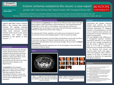
CT angiogram of abdomen and pelvis was concerning for a cecal mass, with wall thickening, irregularity and infiltration of adjacent fat and nodes.
He improved with fluids, antibiotics, and a brief course of vasopressor. His pain recurred with minimal rectal bleeding. Repeat CT abdomen and pelvis 48 hours later was stable. Given the concern for malignancy on imaging, colonoscopy was performed and showed a severely friable and edematous mucosa with ulcerated mass like appearance of a portion of the cecum. Biopsies showed ulceration, acute and chronic inflammation with granulation tissue and reactive changes favoring IIC.
The patient improved with supportive care. Metoprolol and Sacubitril/Valsartan doses were lowered on discharge.
Discussion: Decreased colonic perfusion following exertion on a strong antihypertensive regimen may have predisposed this patient to CI. Typically, IIC presents with bowel necrosis requiring surgery and therefore suggest a more severe presentation and guarded prognosis. Our case was unique in that ischemia in the cecum was localized to one large area, rather than the entire cecum, suggesting involvement of isolated branches of the ileo-colic artery with a milder presentation. While guidelines recommend approaching any case of IRCI as severe CI upfront due to the poor prognosis of IRCI regardless of actual disease severity 5, milder cases such as ours may be carefully identified and managed on the hospital floor.

Disclosures:
Jad Mitri, MD1, Dimo Dimitrov, MD1, Manish Tandon, MD1, Sandeep Krishnan, MD, PhD2. P1980 - Isolated Cecal Ischemia Presenting as a Mass: A Case Report, ACG 2024 Annual Scientific Meeting Abstracts. Philadelphia, PA: American College of Gastroenterology.
1St. Elizabeth's Medical Center, Boston University School of Medicine, Boston, MA; 2St. Elizabeth's Medical Center, Boston University School of Medicine, Brighton, MA
Introduction: Isolated right-sided colonic ischemia (IRCI) is less common than colonic ischemia (CI) typically involving watershed areas in the left colon and entails worse outcomes. Isolated ischemia of the cecum (IIC) is rare, almost solely described in case reports as cecal necrosis.1 2 3 4
Case Description/Methods: The patient is a 68-year-old male with complex cardiovascular comorbidities on Sacubitril/Valsartan and Metoprolol, pulmonary embolism on Warfarin, with screening colonoscopy 3 months prior to presentation when a small tubular adenoma was removed.
He presented with 24 hours of epigastric pain, later diffuse, worse in the right lower quadrant, exacerbated by exertion and meals, and relieved after emesis and a loose non-bloody bowel movement. He had performed strenuous yard work the day prior.
He was afebrile, hypotensive, with diffuse abdominal tenderness, worse in the right lower quadrant, without guarding or rebound tenderness. Labs showed leukocytosis 14,400/μL, INR 2.3, lactic acid 1.4 mmol/L, normal lipase and hepatic panel.
CT angiogram of abdomen and pelvis was concerning for a cecal mass, with wall thickening, irregularity and infiltration of adjacent fat and nodes.
He improved with fluids, antibiotics, and a brief course of vasopressor. His pain recurred with minimal rectal bleeding. Repeat CT abdomen and pelvis 48 hours later was stable. Given the concern for malignancy on imaging, colonoscopy was performed and showed a severely friable and edematous mucosa with ulcerated mass like appearance of a portion of the cecum. Biopsies showed ulceration, acute and chronic inflammation with granulation tissue and reactive changes favoring IIC.
The patient improved with supportive care. Metoprolol and Sacubitril/Valsartan doses were lowered on discharge.
Discussion: Decreased colonic perfusion following exertion on a strong antihypertensive regimen may have predisposed this patient to CI. Typically, IIC presents with bowel necrosis requiring surgery and therefore suggest a more severe presentation and guarded prognosis. Our case was unique in that ischemia in the cecum was localized to one large area, rather than the entire cecum, suggesting involvement of isolated branches of the ileo-colic artery with a milder presentation. While guidelines recommend approaching any case of IRCI as severe CI upfront due to the poor prognosis of IRCI regardless of actual disease severity 5, milder cases such as ours may be carefully identified and managed on the hospital floor.

Figure: Image A- CT angiogram of the abdomen and pelvis showing a possible cecal mass with wall thickening and irregularity as well as infiltration of the adjacent fat with a few nodes.
Images B and C- Inpatient colonoscopy showing a severely friable and edematous mucosa with ulcerated mass like appearance of a portion of the cecum
Images B and C- Inpatient colonoscopy showing a severely friable and edematous mucosa with ulcerated mass like appearance of a portion of the cecum
Disclosures:
Jad Mitri indicated no relevant financial relationships.
Dimo Dimitrov indicated no relevant financial relationships.
Manish Tandon indicated no relevant financial relationships.
Sandeep Krishnan indicated no relevant financial relationships.
Jad Mitri, MD1, Dimo Dimitrov, MD1, Manish Tandon, MD1, Sandeep Krishnan, MD, PhD2. P1980 - Isolated Cecal Ischemia Presenting as a Mass: A Case Report, ACG 2024 Annual Scientific Meeting Abstracts. Philadelphia, PA: American College of Gastroenterology.
